CLSM Confocal laser scanning 3D optical micrographs of Tasmanian tiger hair
Introduction
A series of scanning electron micrographs of Tasmanian tiger hair (Thylacinus cynocephalus, also called "thylacine") was produced in 2017 by Rehberg (author), in collaboration with the Commonweath Scientific and Industrial Research Organisation (CSIRO). Prior to this research, thylacine hair imaging was limited to grainy black and white light microscope micrographs (Taylor, 1985) and hand drawn illustrations (Lyne & McMahon, 1950).
Lyne and McMahon identified three types of hair in the thylacine coat, in contrast to all other species in the family Dasyuridae occurring in Tasmania, which have two. Taylor presented a dichotomous key for distinguishing thylacine hair from other mammals found in Tasmania.
The hair fibres used in the scanning electron microscope (SEM) work were acquired through purchase from a dealer in antique microscope equipment and specimens and dated to between 1850 and 1890. The SEM research, together with the earlier publications, confirmed the hairs to be from a thylacine and all three Tasmanian tiger hair types were present and imaged. The resulting scanning electron micrographs and further information about the context of that research and provenance of the hair sample are given in SEM Scanning electron micrographs of Tasmanian tiger hair.
Subsequent to the SEM research, CSIRO retained hair fibres for the purpose of research using additional imaging techniques. One of these is confocal laser scanning microscopy (CLSM), which produces pseudo three dimensional (3D) optical micrographs. The full set of the CLSM 3D optical micrographs is presented here.
Technique
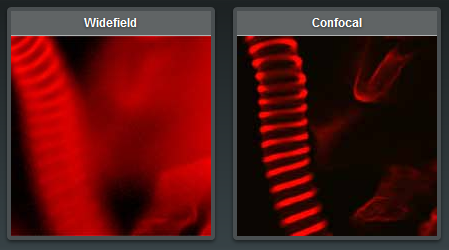
In simple terms, the key concept behind CLSM is that the microscope focuses only on an ultra-thin section of the subject sample at any given time. The benefit gained is that CLSM produces images of far better resolution and contrast than can be achieved through light microscopy. Finally, for thick samples such as the thylacine hair in this study, the system scans multiple sections in sequence, one at a time, and these sections are then merged together to present a single final image of the entire sample. The final image is higher resolution and shows better contrast than can be acheived using a light microscope. It also shows a complete focus throughout the specimen's depth (leading to the "3D" description) which cannot be acheived with a light microscope. However, similarly to a light microscope, and differently to the SEM research, the CLSM images are in colour.
An example of the difference in focal depth, resolution and contrast between light microscopy (labelled "Widefield", here) and confocal laser scanning microscopy is illustrated by Nikon (Microscopyu, 2016) and reproduced below. (This is not thylacine hair.)
Procedure
The thylacine hair fibres were placed on a flat surface and imaged using an Olympus OLS 4100 LEXT by Colin Veich and Mark Greaves of CSIRO.
Images
Below are the first published confocal laser scanning microscope images of Tasmanian tiger hair. These are also the highest resolution true colour images of thylacine hair published to date. These micrographs illustrate the hair surface morphology, and in particular the scale patterns are apparent.
Two scans were performed of each hair type: outer; guard and under. A composite image of the three hair types at the same scale is also presented.
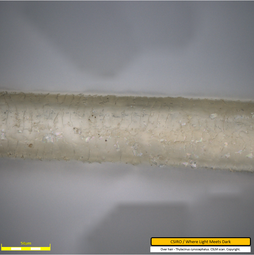
Over hair CLSM micrograph 1
This micrograph shows a Tasmanian tiger over hair fibre with scale bar at 50 micrometres. The scale pattern is visible as a series of darker lines traversing the fibre. Dirt particles are visible as darker spots adhered to the fibre. The top edge of the fibre shows how scales to the left overlap scales to their immediate right, suggesting the hair root is toward the left of frame.
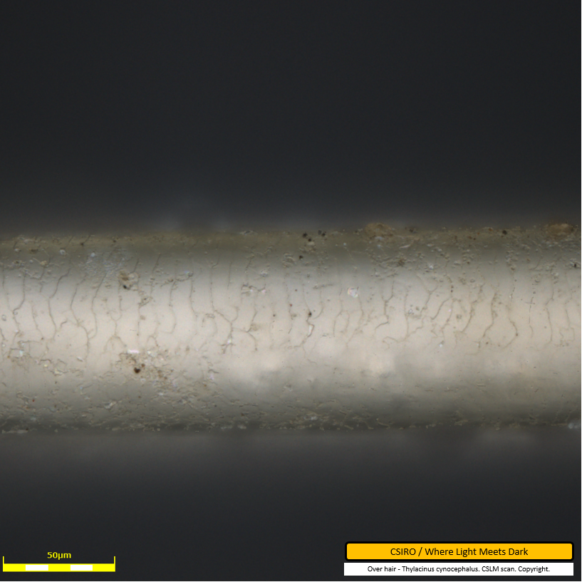
Over hair CLSM micrograph 2
The cuticle scale pattern on a thylacine over hair is visible in this micrograph as a series of lines traversing the fibre. Dirt particles are seen as darker spots adhered to the fibre.
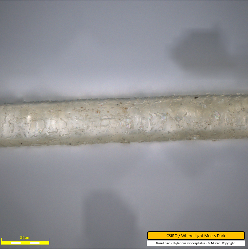
Guard hair CLSM micrograph 1
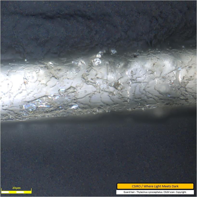
In this micrograph, the scale pattern on a guard hair is visible as dark lines traversing the fibre along the centreline of the fibre, and as pale lines just below the top edge of the fibre. Along the top edge in particular, the scales can be seen overlapping one another. Scales to the left overlap adjacent scales to their right suggesting the root end of the hair is off-frame, to the left. Some dirt particles are visible as darker spots adhered to the fibre.
Guard hair CLSM micrograph 2
This enlarged micrograph (scale bar 20 micrometres, compared to 50 micrometres for prior images) shows the scale pattern of a guard hair in detail. Apparent damage to the cuticle (outer layer) near the bottom edge of the fibre is visible in the left half of the image. Some dirt particles are also visible in this image, adhered to the fibre.
Under hair CLSM micrograph 1
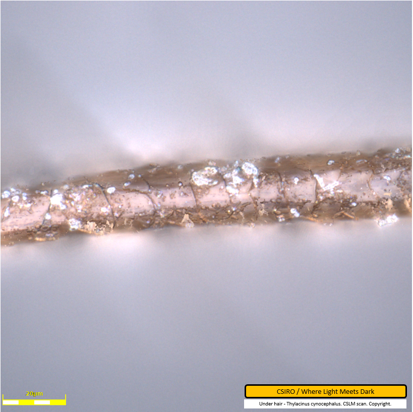
This micrograph of thylacine under hair is also at an enlarged scale, having a scale bar of 20 micrometres. Under hair is considerably thinner than over and guard hair. The scale pattern is clearly visible traversing the fibre. Some dirt particles are adhered to the fibre also. It can be seen at a few points that scales toward the left overlap scales immediately to the right, suggesting the root of the hair is off-frame to the left.
Under hair CLSM micrograph 2
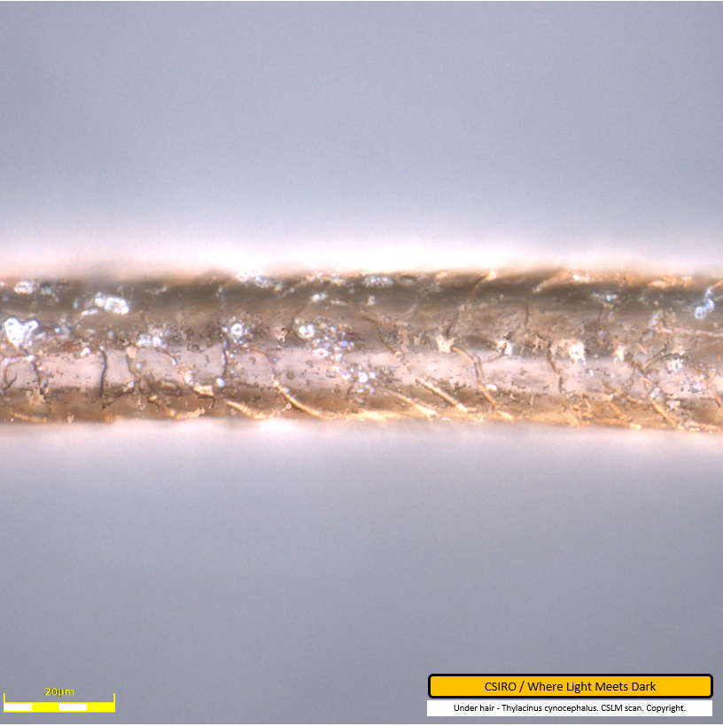
This micrograph shows thylacine under hair with a 20 micrometre scale bar. The cuticle scale pattern is clearly visible as lines traversing the hair fibre. The root of the hair is off-frame toward the left. This image has been captured at the same enlargement as the prior image of the same fibre. However in this image, the fibre is considerably thinner at the left edge of the image, ie. toward the root. This decrease in hair thickness toward the root was observed during the scanning electron micrography also. Due to the small number of fibres analysed, it is not known how common this trait is. Dirt particles are also visible adhered to this fibre.
Composite CLSM micrograph - over, guard and under hair to scale
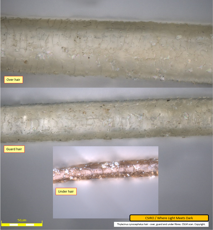
The image below is a composite produced from three of the images above. The over and guard hairs were captured at the same scale as each other during micrography. The under hair was captured at a different scale. The under hair image was reduced in size in order to represent it at the same scale as the other two hairs. As such, the three thylacine hair types are presented here, side by side, to the same scale.
This image is interesting as it provides a photographic example of the differing scale patterns as presented by Lyne & McMahon as an illustration. The scales on the over hair form narrow bands that traverse the fibre. The scales on the under hair are approximately equal in length (measured along the fibre) but do not appear as narrow due to the hair width being much reduced in comparison to the over hair. The scale pattern on the guard hair appears less banded and more erratic.
Further research and acknowledgements
This scanning work was carried out by CSIRO under a generous offer of support in response to ABC News coverage of Where Light Meets Dark's crowd funding campaign to produce high quality light microscopy images of Tasmanian tiger hair. The author would like to acknowledge the generosity of Colin Veitch and Mark Greaves, CSIRO Manufacturing - Waurn Ponds and Clayton Laboratories respectively - and the coverage of ABC.
The stated goal of the crowd funding project is to produce light microscopy images of Tasmanian tiger hair that may become a reference base against which researchers may compare samples collected in the field for the purpose of a potential thylacine match. The confocal laser scanning micrographs presented here present a level of resolution and clarity that is not possible using widefield light microscopy and exceed the clarity expected for the reference base. While most field researchers will not have access to CLSM equipment with which to check fibres collected in the field, these CLSM images will still be useful for comparison with light microscopy images that a field researcher may produce relatively inexpensively.
The CLSM images presented here illustrate surface morphology and scale patterns on the three thylacine hair types. The completed light microscopy work will additionally illustrate internal fibre structures, namely the presentation of the medulla and pigment throughout the hair fibres.
The author would also like to acknowledge the many backers of the light microscopy project, and the project's sponsor, Dr Stephen Sleightholme, without whom there would not have been news coverage and the generous offer from CSIRO for the microscopy.
Citing this article
Rehberg, C. (2017). CLSM Confocal laser scanning 3D optical micrographs of Tasmanian tiger hair, Accessed: date, http://www.wherelightmeetsdark.com.au.
References
- Lyne, AG and McMahon, TS (1950) Observations on the surface structure of the hairs of Tasmanian monotremes and marsupials. Papers and Proceedings of the Royal Society of Tasmania. pp. 71-84. ISSN 0080-4703
- Microscopyu (2016) Laser Scanning Confocal Microscopy, Accessed: 9 June 2017, https://www.microscopyu.com.
- Rehberg, C. (2017) SEM Scanning electron micrographs of Tasmanian tiger hair, Accessed: 9 June 2017, http://www.wherelightmeetsdark.com.au.
- Taylor, Robert J (1985) Identification of the hair of Tasmanian mammals. Papers and Proceedings of the Royal Society of Tasmania 119, 1985. pp.69-82. 0080-4703.
