OLM Optical Light micrographs of Tasmanian tiger guard hair
This is one page of results for the article OLM Optical Light micrographs of Tasmanian tiger hair.
To cite this page of results or any image within this page, see "Citing this article" at OLM Optical Light micrographs of Tasmanian tiger hair.
This page contains 52 images totaling 29MB.
Notes
Although the thylacine has three hair types in its coat, the guard hair appears to be the type given most consideration by Lyne & McMahon and Taylor. Taylor's key rests solely on working with the guard hair.
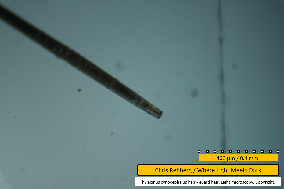
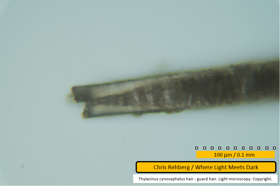
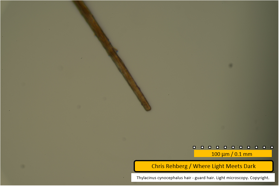
File 2980 - proximal end
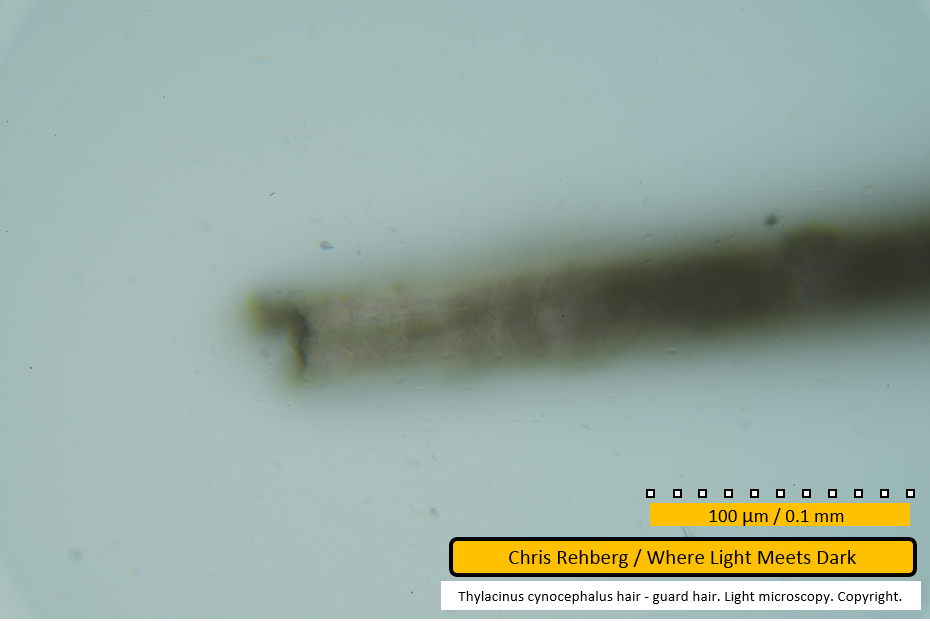
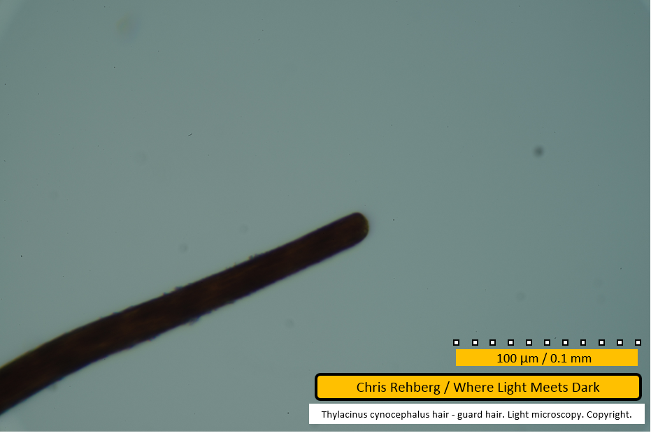
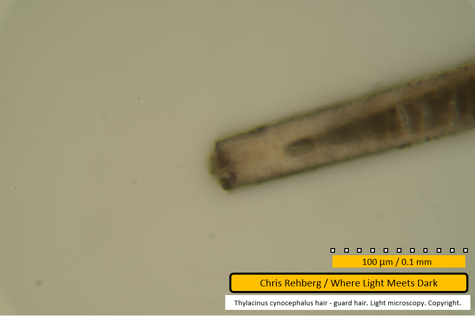
An end of the guard hair is shown here. Faint pale lines traversing the fibre reprsent the scale pattern. The medulla gives the fibre its dark core almost to the very end. The end is abruptly tapered and apparently cut. At the very tip the medullary core constitutes less than half the diameter of the fibre - in contrast to the remainder of the fibre in this image where it constitutes almost 100% of the fibre's width. No conclusion can be drawn about this being the distal or root end of the fibre based solely on the tapered tip and the relatively thinner medulla. Lyne and McMahon note "the majority of hair shafts [across all of the mammals considered and not just the thylacine] show some variation in diameter throughout their length. The base is usually of less diameter than the mid-region. In most of the Tasmanian marsupials the protective hairs are widest distal of the mid-region and the tip usually tapers off to practically nothing" (pp 75-6). As such, neither the root nor tip is the thickest part of the fibre and hence in both cases may appear tapered. A definitive conclusion may be drawn by examination of the scale pattern.
No images of the present fibre (seen in file 2980) are adequate to discern sufficient detail regarding scale pattern, however files 2997 and 2999 - 3001 show near-identical features in a separate guard hair (the third examined, chronologically, and the third presented on this page) and image 3002 shows sufficient detail regarding that guard hair's scale pattern to conclude that in both cases the proximal end is being examined (ie here, in file 2980 and for the other guard hair, in files 2997 and 2999 - 3001).
Further, file 2986 shows an end of yet another guard hair (the second examined chronologically, and the second presented on this page) at 4x greater magnification than used here (ie. captured at 400x, compared with 100x here) that is markedly thinner than the one seen here. As such, file 2986 perhaps better fits the description of tapering "off to practically nothing". This supports the conclusion that the present image shows a proximal end while image 2986 shows a distal end. On the basis of the guard hairs examined in the present study, the proximal end of a guard hair may be distinguished from the distal end on the basis of medullary appearance and overall thickness of the fibre - assuming the fibre is not prematurely terminated at either end. That is, for guard hairs it may not be essential to examine the scale pattern in order to distinguish the root and distal ends.
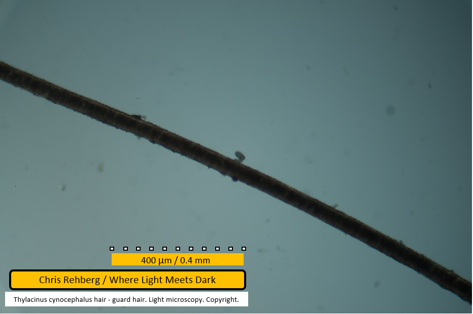
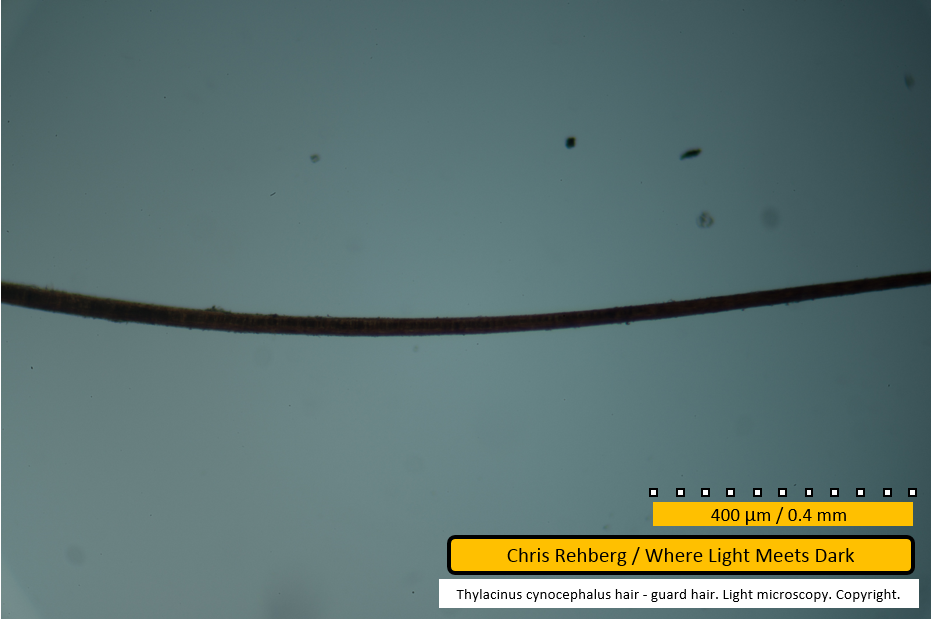
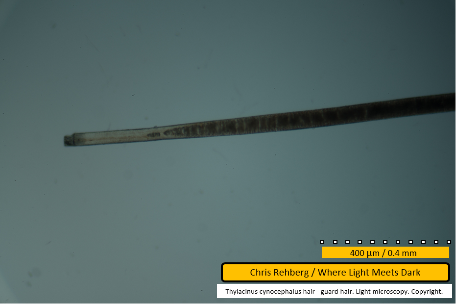
This, and subsequent images showing the 400um scale bar, were captured at approximately 100x enlargement. When compared with the proximal end (file 2948) or distal end (file 2949, lower fibre, which is more intact) of the under hair (both images being at same scale), it is clear the guard hair diameter is approximately an order of magnitude thicker than in the under hair. The vertical line at right of frame is the edge of the cover slip and unrelated to the fibre.
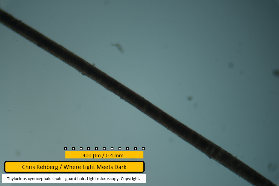
File 2981 - mid section
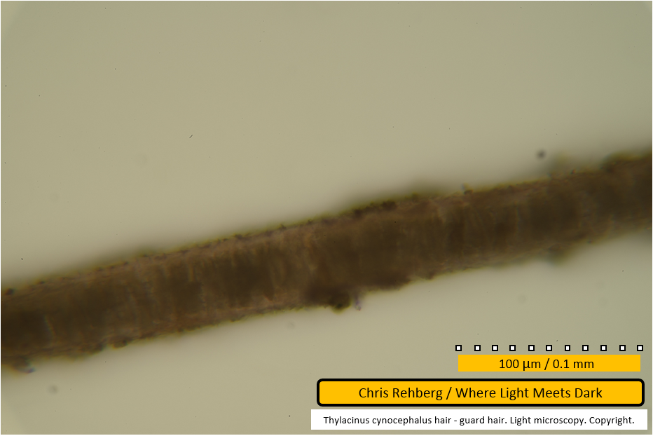
The same fibre, panning left. Again the dark medullary core dominates. Minor spacing, or vacuolation, is evident between medullary cells at lower right.
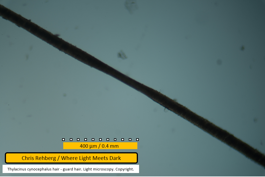
File 2982 - mid section
In this frame there is a notable thinning of the fibre at centre. In the earlier SEM work it was assumed this is due to distortion caused by manipulation of the fibre using tweezers. While Lyne & McMahon note that a fibre's diameter can vary over its length (p 76), the natural variance is unlikely to be as abrupt as seen here. However Lyne & McMahon also note that cross sectional shape may vary along a fibre's length (p 76). Taylor notes the thylacine's guard hair cross section is primarily oblong (see key, p 69). By inspection during this study, whilst varying the focal depth under the light microscope, some fibres gave the appearance of having a compressed shape (ie. as expected for oblong cross section) that may be twisted along its length - comparable to a twisted party streamer. Such a twist would have an appearance similar to that seen here when captured as a still frame. As such, the exact cause of apparent compressions such as this remains unverified.
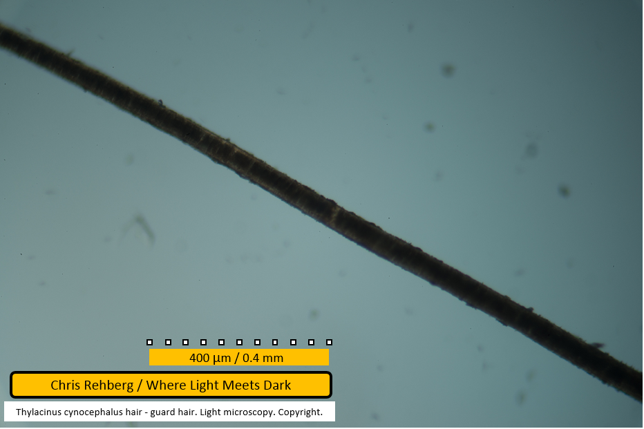
File 2983 - mid section
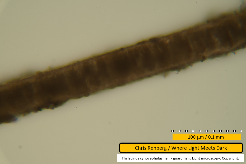
Further along the same fibre. Dirt particles are seen adhered to the fibre along the edges. The appearance of the medulla has been consistent through recent images.
File 2984 - mid section
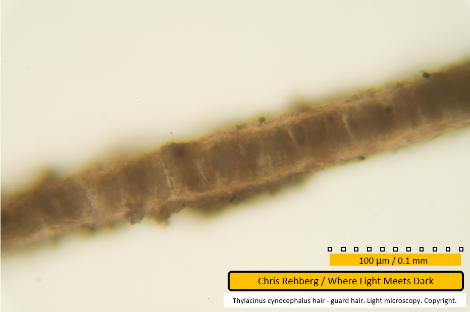
Further along the same fibre. Again, the appearance is consistent with prior images in the sequence.
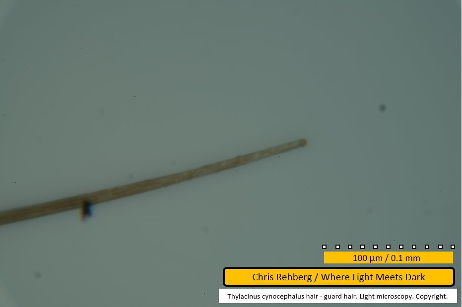
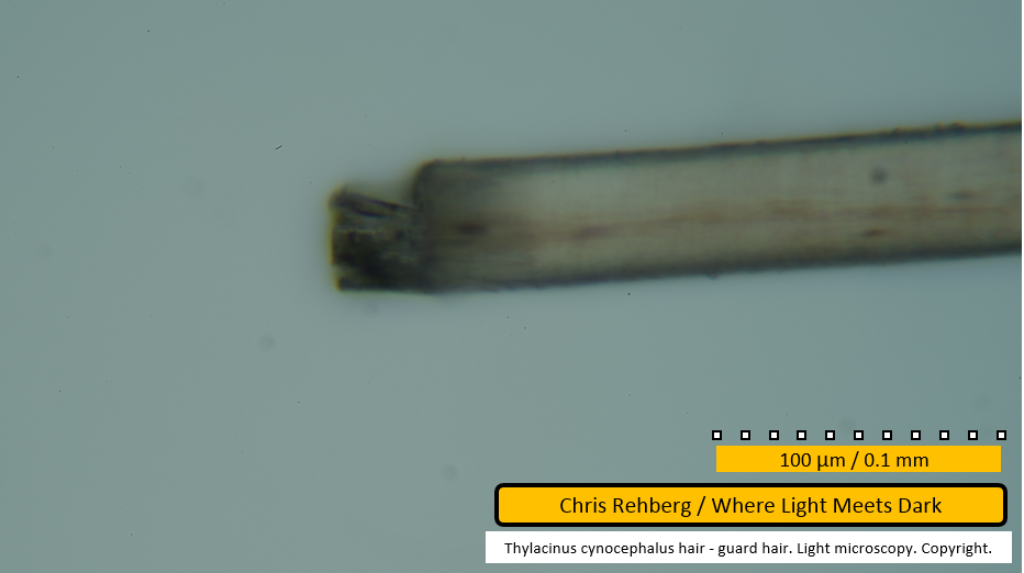
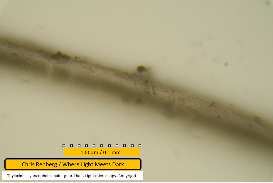
File 2986 - distal end
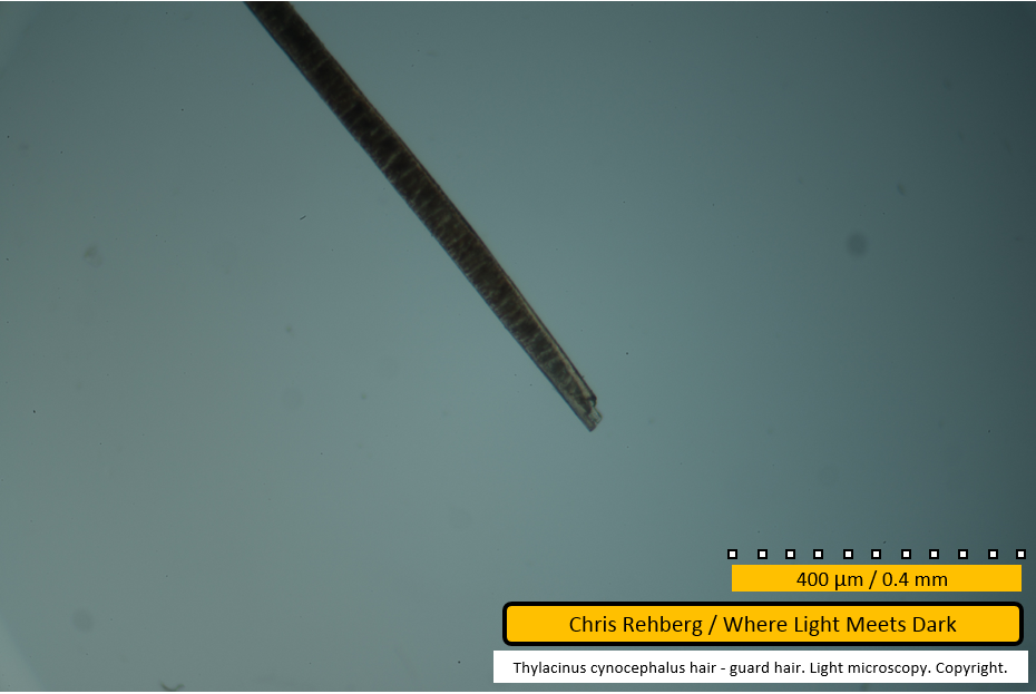
Noting the comments on file 2980 (which shows a different fibre), it is concluded the present image (file 2986) shows the distal end of a guard hair. Note the absence of the medulla for considerable distance (in contrast with the root end as seen in file 2980 - but see also file 2997 - and) in common with the distal ends of under hairs, such as seen in file 2978.
There is an apparent longitudinal striation of paler colour than the predominant straw colour, at left of frame, but the cause of this is unknown.
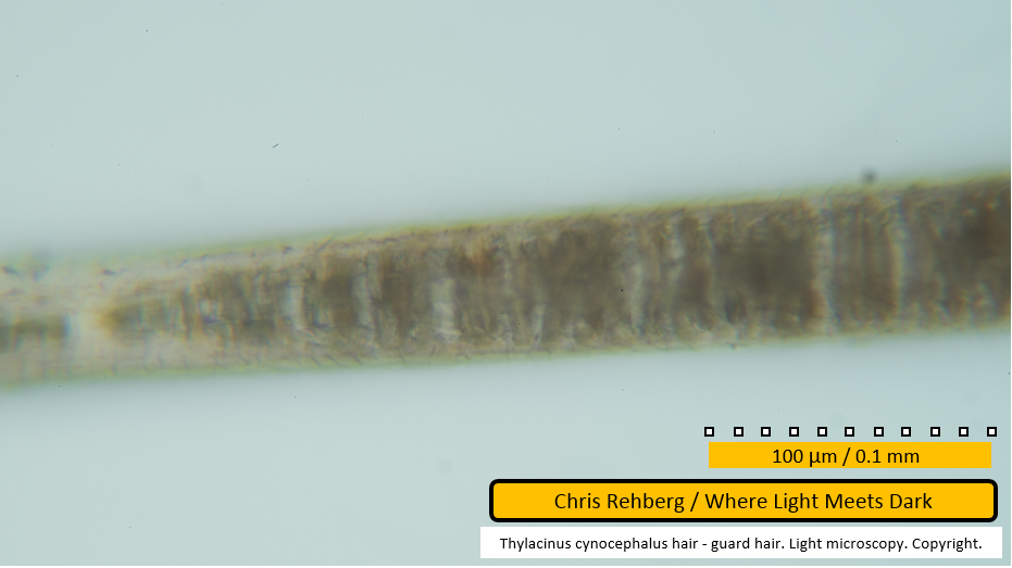
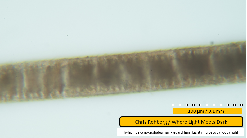
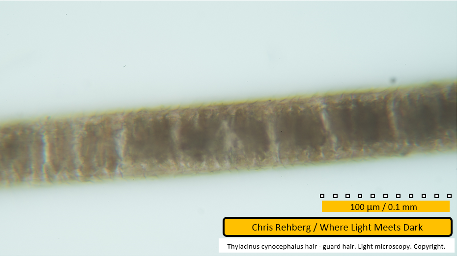
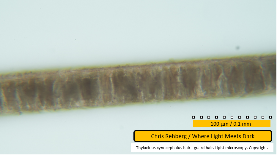
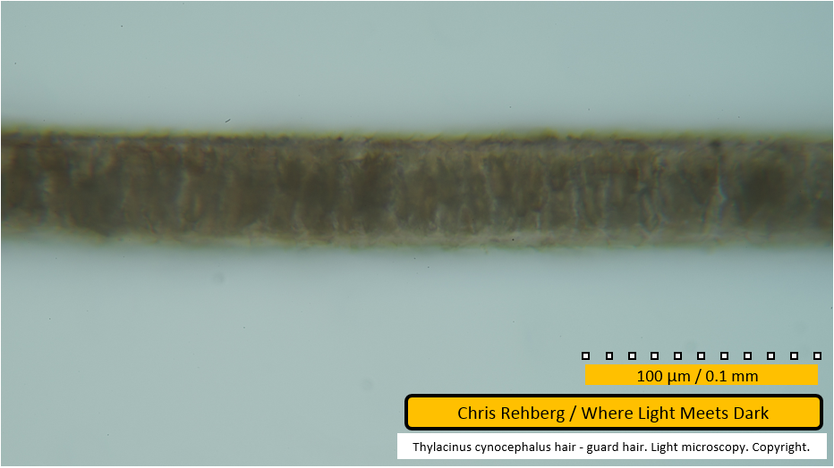
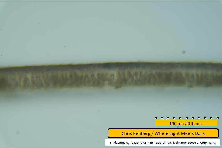
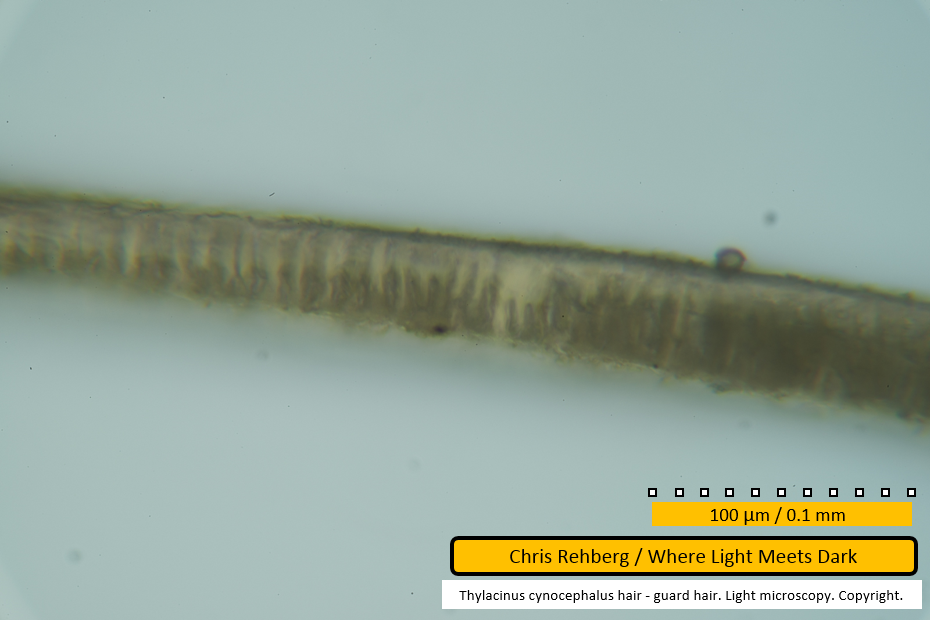
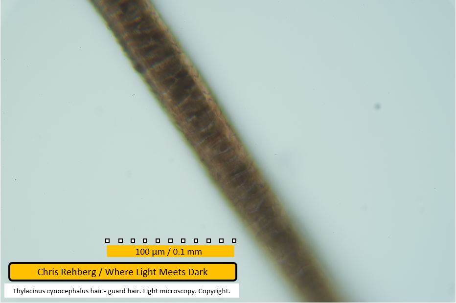
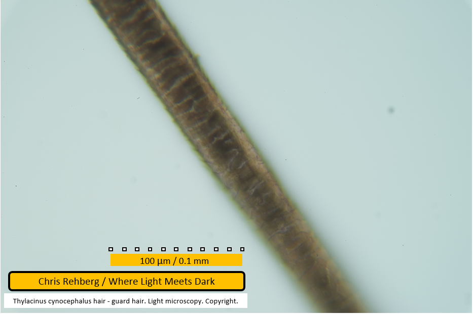
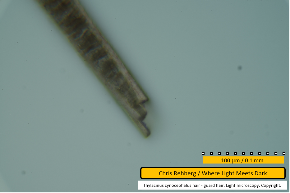
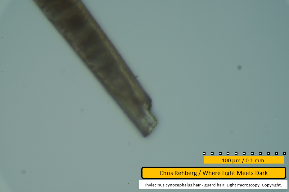
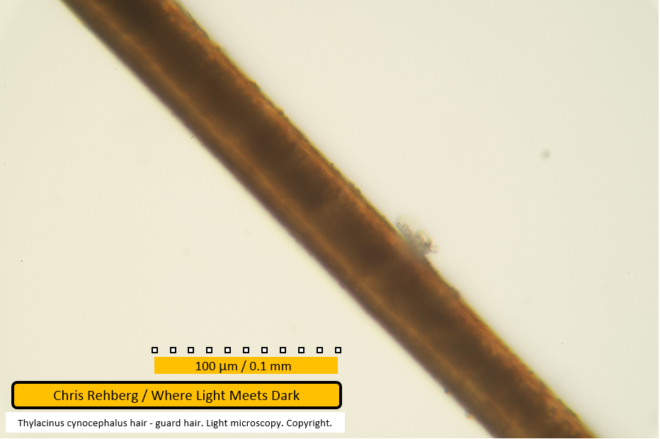
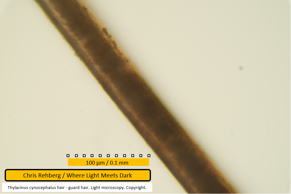
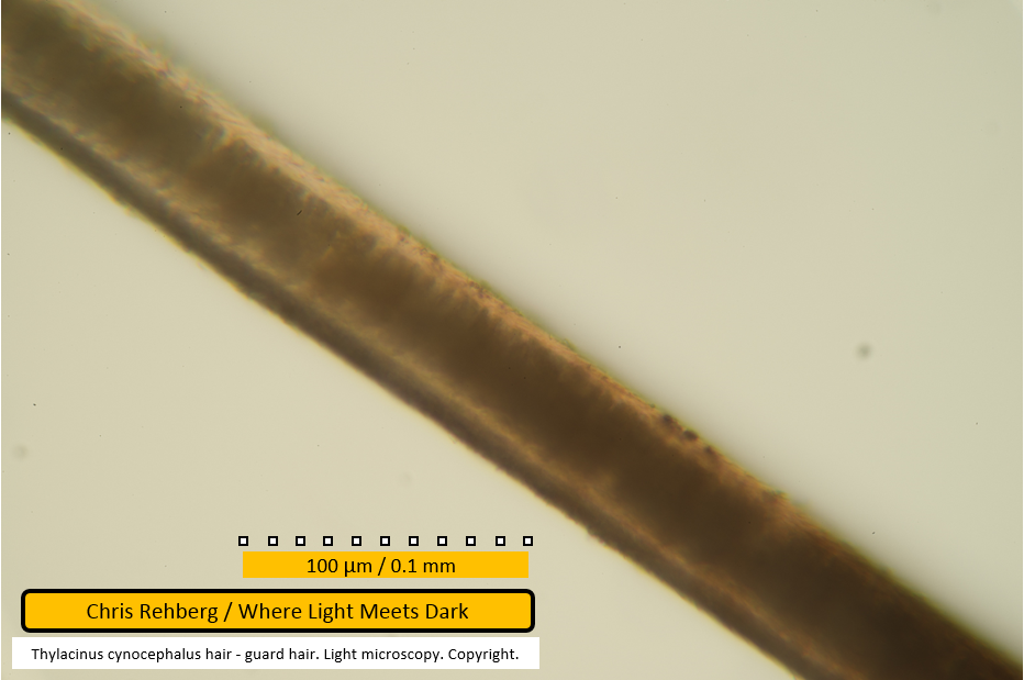
This, and subsequent images showing the 100um scale bar, were captured at approximately 400x magnification.
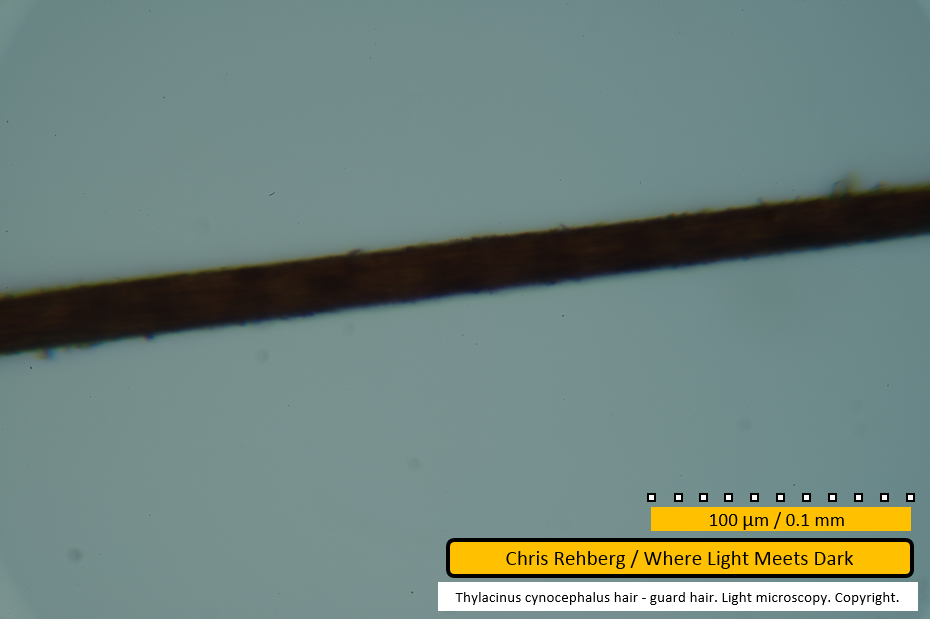
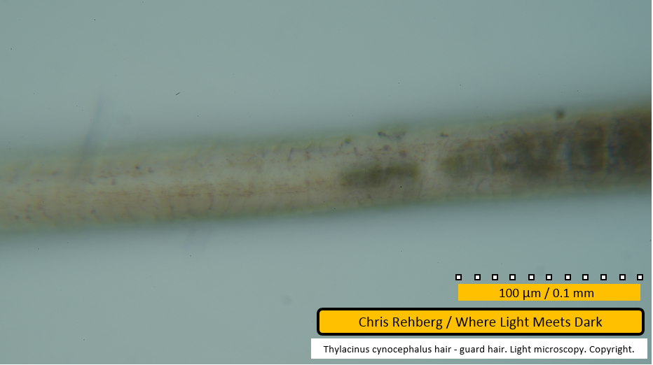
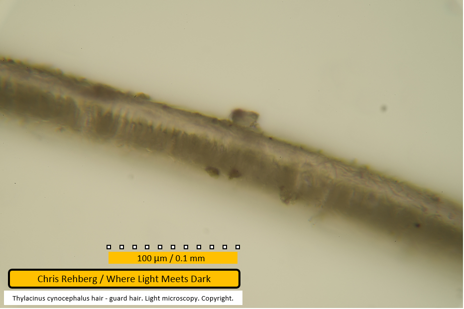
File 2987 - mid section
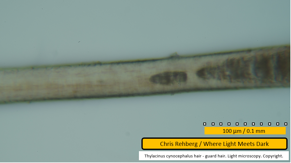
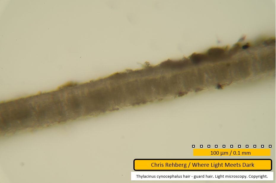
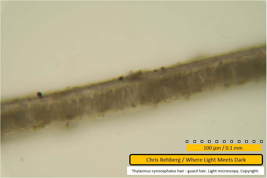
Considerably further along the fibre, the diameter is much thicker. Note the apparent thick, darker, transverse banding. This is not due to the medulla which, in comparison, can be seen in the next image (file 2988). In contrast, the medulla initially traverses less than 50% of the diameter of the fibre and has much narrower spacing between medullary cells than seen between bands in this image (file 2987). The bands visible here are due to pigmentation. In other words, the distal end of thylacine guard hairs is striped!
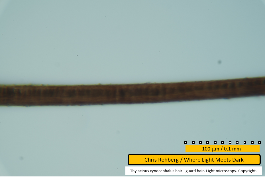
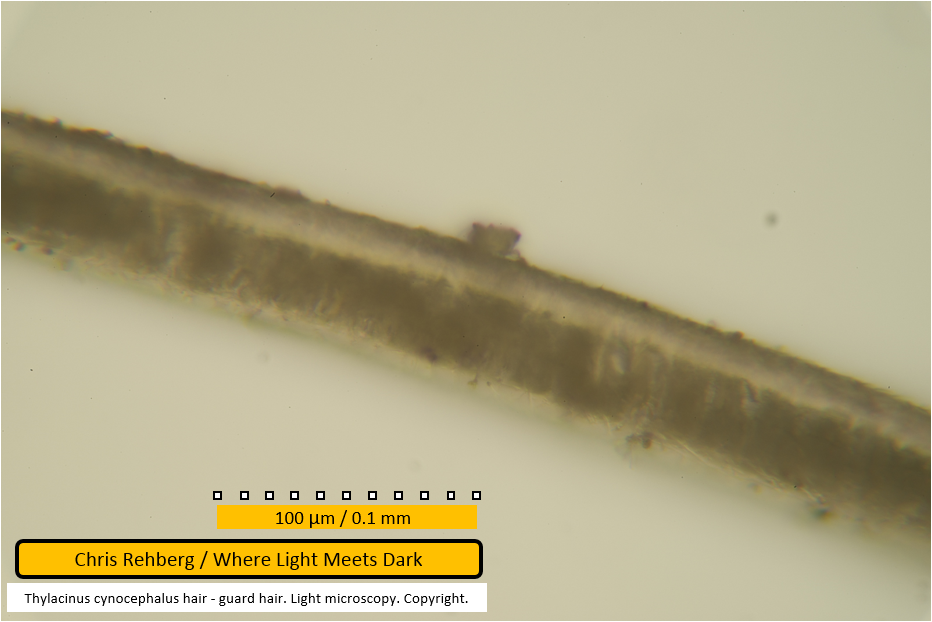
File 2988 - mid section
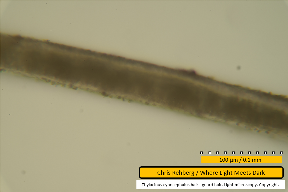
The medulla becomes apparent here as a uniserial ladder initially traversing less than 50% of the diameter of the fibre. As we progress our examination of the fibre toward the left, the medulla thickens and occupies a greater proportion of the width of the fibre.
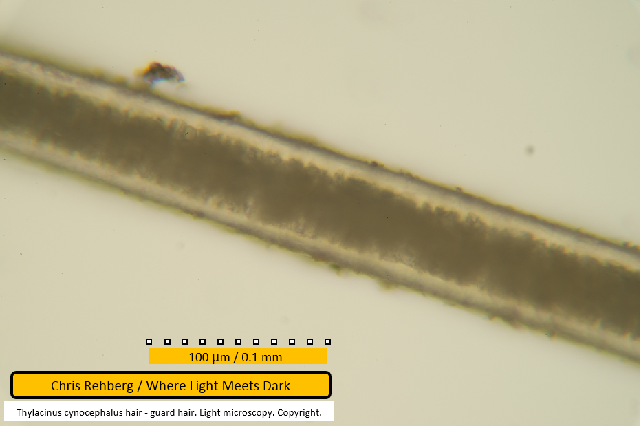
File 2989 - mid section
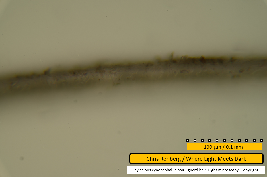
Note the reduction in magnification for this image. At this reduced magnification the medulla is still apparent, thicker toward the left of frame.
File 2992 - proximal end
Returning to the higher magnification (captured at 400x), this shows the other end of the same fibre as shown in the immediately prior images. Focus is on the fibre surface where the scale pattern can be seen as very faint pale or dark lines traversing the fibre in imbricate fashion.
File 2993 - proximal end
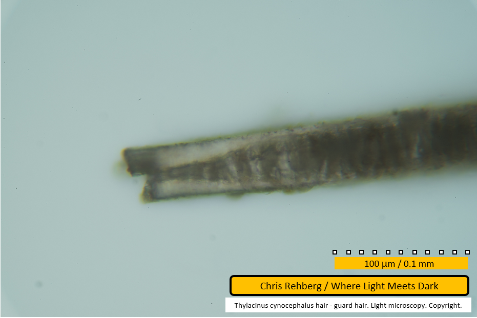
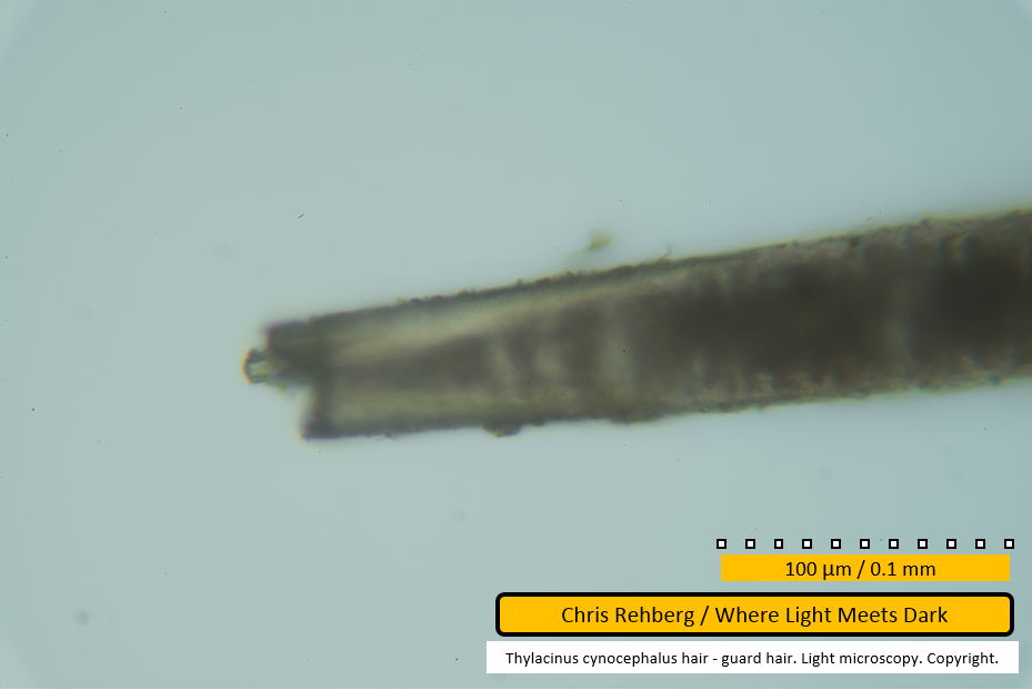
This shows the same fibre end but with the focal depth adjusted to clarify the internal structure (as opposed to the surface scales seen in file 2992). The overall shape of the taper here is similar to that found in file 2980 which showed a different guard hair. File 2980 was concluded to be the root of that guard hair on the basis of comparison with a third guard hair showing similar features and sufficient detail to observe the lie of the scale pattern (see files 2997 and 3002). That being the case, it is concluded that this image (file 2993) also shows a root end - in this case, of this second guard hair.
Note the rapid thinning of the medulla. In fact the medullary cells rapidly grow at this point with the hair root toward left and the more mature medullary cells toward right.
In the present case the root end appears irregularly broken. This image, the prior image and the following two images all show the same root but at varying focal depths, to give a sense of the appearance of this irregular break.
File 2994 - proximal end
See notes on files 2992 and 2993.
File 2995 - proximal end
See notes on files 2992 and 2993.
File 2997 - proximal end
This image shows a third guard hair fibre. A subsequent image (file 3002) shows sufficient detail in the scale pattern to conclude this is the root end of this fibre. The fibre is thicker where the medulla is present (at right), then tapers where the medulla thins, then remains thin at the end where the medulla is absent. The tapering appearance of the fibre at the point where the medulla thins and ends, together with the rapid thinning of the medulla, is comparable with the appearance of the two prior guard hair fibres at their proximal ends (as seen in files 2980 and 2993). The conclusion drawn is that the ends examined for those two fibres (in files 2980 and 2993) are also proximal ends.
In this image (file 2997), the portion of fibre lacking a medulla is notably longer than that seen in the two prior fibres. It is not known whether or not the medulla-free portion at the root end of guard hair fibres is of relatively consistent length between fibres. Natural variation may be due to the age of the fibre, the location on the body from which the fibre was sourced (although in this study it seems likely all hairs were sourced from a single location on the pelage) or other unexplained reasons. However if this length is typically consistent between fibres, this would suggest the prior two fibres (and the following two fibres) have been cut some distance from the actual terminus of the fibre, whereas this fibre has been cut or broken nearer to the actual root.
This image has been captured at lower magnification than the root of the second guard hair fibre examined in the prior four images. Subsequent images are captured at higher resolution and provide direct comparison between this and the prior fibre.
In this image a single brownish-orange striation is seen where there is no medulla. This author speculates the cortex may contain a central hollow at this point in which the medullary cells grow - as this striation aligns with the commencement of the medulla, at least in this view of the fibre.
File 2999 - proximal end
The broken or cut end of the fibre, likely near the natural terminus close to, or within the follicle. This broken end has a greyish-black appearance with cause unknown. The brownish-orange striation discussed under file 2997 is seen magnified here. Small inclusions, called ovoid bodies, are visible in the cortex as brown or dark spots.
File 3000 - mid section
This image shows the commencement of the medulla, which is shown out of focus. The focal depth emphasises the fibre surface scale pattern.
File 3001 - mid section
This image shows the same region as file 3000 but with focus on the medullary core rather than the surface scale pattern.
File 3002 - mid section
The break in the medulla seen in the prior image (file 3001) is seen here at the far left of frame. The scale pattern is visible along the edges of the fibre and suggests that the edge of an individual scale that is toward the right of frame is lifted further away from the fibre than the edge of the same scale that is toward left of frame. This confirms the root of the fibre is toward left of frame.
File 3003 - mid section
The "lifted" scales along the fibre edge again confirm the root to be toward the left. The medulla spans almost the full width of the fibre. The broad bands between medullary cells are vacuolated spaces. The scale pattern is not visible traversing the fibre as the fibre surface is not in focus.
File 3004 - mid section
Continuing the same fibre, showing similar features.
File 3005 - mid section
Continuing the same fibre, showing similar features.
File 3006 - mid section
Continuing the same fibre, showing similar features.
File 3007 - mid section
Continuing the same fibre. The medulla takes on an unusual appearance in this frame - seemingly visible only along the lower half of the fibre. The cause for this is unknown and it is unknown whether the medulla would have had this appearance in life - or whether this may be an indication of desiccation or decay over time at this point in the fibre. There remains also a possibility that the appearance is an artefact of light (without any medullary non-conformity) due to, for example, physical deformity of the fibre at this point through, for example, handling with tweezers or otherwise. See, however, notes on the next image (file 3008).
File 3008 - mid section
The atypical appearance of the medulla discussed in the prior image (file 3007) is visible again in the left of frame here. At right of frame the typical medullary pattern is again seen. The fibre width appears narrower at left of frame and may indicate a correlation with the atypical appearance of the medulla. However the fibre edge is out of focus at left of frame and this can affect the apparent width of the fibre. An accurate assessment of the fibre's width - and whether any gross deformity exists - can be made by observing the fibre as the depth of focus is changed throughout the fibre's depth. If the fibre is found to actually be thinner at the point where the the medullary appearance is atypical, this would suggest the medullary cells are in fact reduced in size at this point; for example, compare with any of the guard hair proximal end images that show rapid thinning of the fibre width in correlation with reduction and absence of the medulla.
Lyne & McMahon note "the marked changes in scale form, which are usually found along a single hair, are considered to be of an inherent and genetic character and are not caused by varying amounts of attrition or by dehydration of the hair shaft". This author proposes that growth of the medullary cells expands the width of the fibre, leading to the spinuous (slightly raised) appearance of the scale pattern where the medulla is present (and hence, explains the imbricate scale pattern near the root where the medulla is absent).
File 3009 - mid section
Few features are notable in this image although the colouration remins sandy.
File 3010 - distal end
The distal end again exhibits broad dark banding over a dark chocolate brown colour as seen in earlier guard hairs. In this case the end is rounded and likely cut, meaning the longer, thinner, straw-coloured (and unbanded) tip is absent.
File 3011 - proximal end
This image shows the root end of the fourth guard hair examined. Based on the earlier analyses on this page, the rapidly tapering medulla combined with reduction in fibre width has proved typical for the proximal end of guard hairs. In common with two of the three prior guard hairs, the fibre is broken at this point where the medulla commences. While the method employed for removing the hair samples from the animal is unknown, the relative consistency of breaks at this point suggest the fibre is weaker near the root at the point where medullary cells form.
File 3012 - mid section
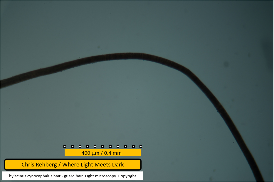
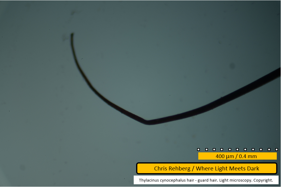
This guard hair, on inspection, was notably bent. This image shows one point where the fibre has been kinked, at low magnification.
File 3013 - distal end
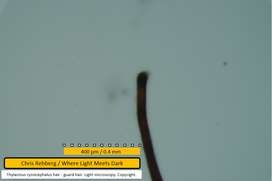
This image shows a second point at which this fibre has been kinked - just prior to the distal end.
File 3014 - distal end
This image shows the distal end at high magnification. The dark banding pattern against a dark brown base is again apparent. The very end of the fibre has a tufted appearance - it is unknown whether this represents dirt adhered to the fibre, a natural fraying of scales or organic growth. Again the lack of a straw-coloured, unbanded tip suggests the end has been broken.
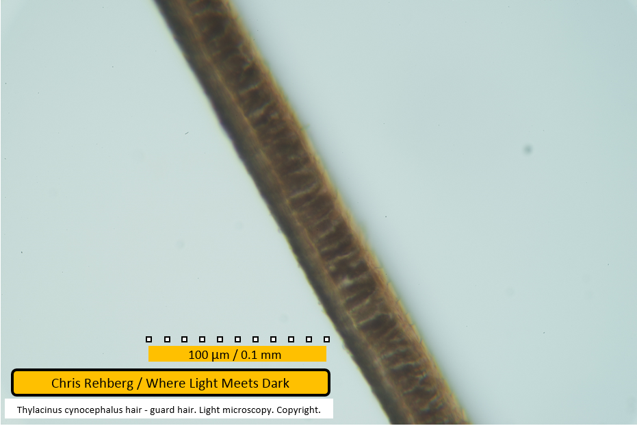
File 3015 - mid section
In this image the same fibre is examined mid-region at higher magnification. This and subsequent images for this fibre attempt to illustrate the surface scale pattern under light microscopy as we approach the root end of the fibre. In this image the scale pattern is visible along the right/upper edge of the fibre
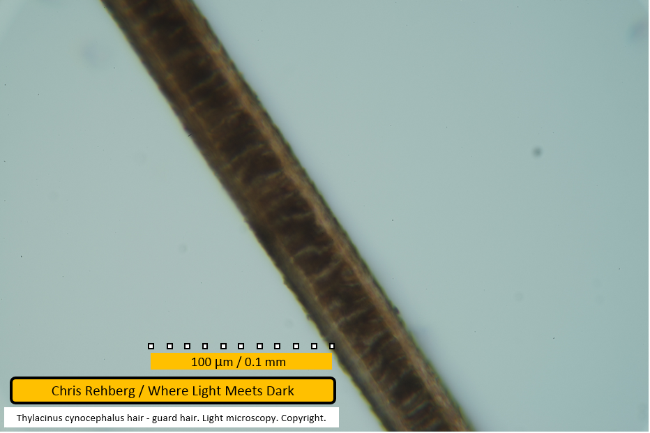
File 3016 - mid section
The same fibre as we pan down and right, toward the proximal end.
File 3017 - mid section
The same fibre clearly exhibiting the scale pattern
File 3018 - mid section
The same fibre showing scales at edges in upper half of frame, traverse scale pattern toward lower half of frame and thinning of the medulla as we scan down the frame.
File 3019 - proximal end
The broken proximal end of this fibre.
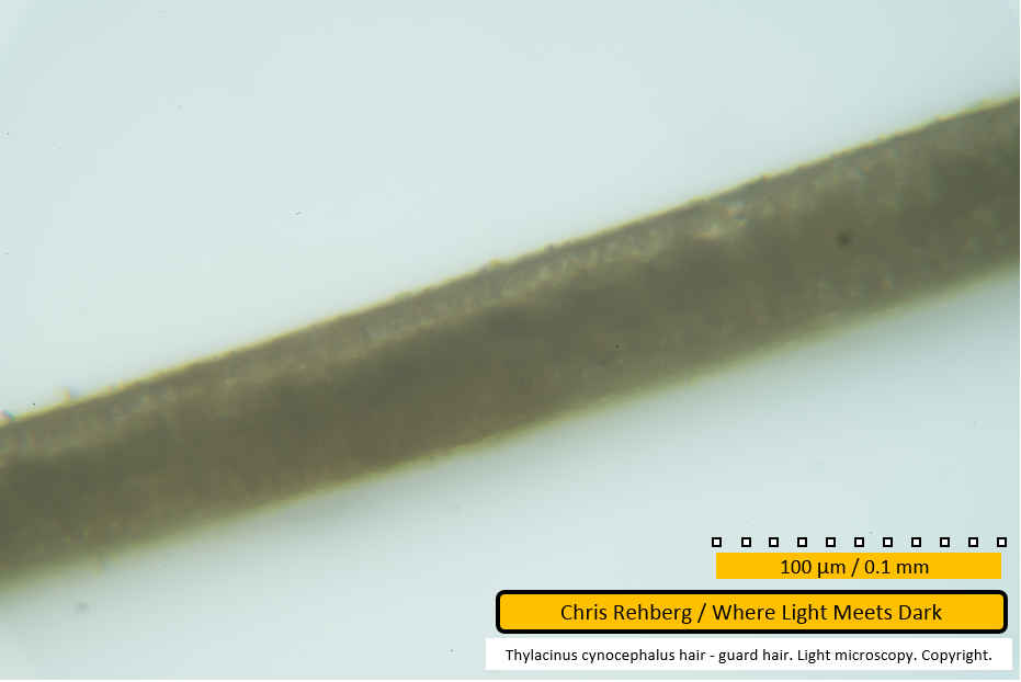
File 3020 - proximal end
This is the same fibre with a different focal point to emphasise some of the features of the shape of the broken end.
File 3022 - proximal end
This image shows the proximal end of the fifth guard hair fibre examined. In common with three of the four prior guard hair root ends, this one is also broken at the point where the medulla commences. The lifted scales are visible along the upper edge and again confirm this thicker end of the guard hair as being the root.
File 3023 - mid section
A considerable amount of dirt is seen adhered to this fibre, particularly on the lower edge at left of frame. The medulla is visible for being notably darker than the remainder of the cortex, but is not sharply in focus.
File 3024 - mid section
Again a considerable amount of dirt is seen adhered to the fibre.
File 3025 - mid section
The same fibre, mid region.
File 3026 - mid section
In this image the medulla can be seen to narrow in the right half of frame. The unusual appearance of the medulla seen in file 3008 also appears in this present fibre and it more notable in the following image (file 3027).
File 3027 - mid section
In this frame the unusual appearance of the medulla resembles that seen in an earlier fibre in file 3008. In this image the medullary cells appear reduced in size and transparent - possibly suggesting dessication. It is unclear whether the cells appeared this way in life or whether this effect occurred post mortem. One speculation is that this sequence of medullary cells failed to develop normally at time of growth and that the underlying cause for this was nutritional and thus affecting all (or majority) hair growth at the same time. This would explain why the same phenomenon is seen in two separate fibres. A re-examination of the fibres in detail may reveal more, or most of the guard hairs affected in this way. It would be an interesting investigation to count the proportion of fibres affected, the distance from medullary commencement where this phenomenon occurs, and a count of medullary cells affected. It is possible this feature testifies to a significant event in the life of the thylacine from which these fibres were taken. If the growth rate of medullary cells in guard hairs were known, the count of affected cells may give an indication of how long this life event affected the thylacine. For example - does it represent a period of poor nutrition due to extraordinary environmental conditions? A period of refusing food during captivity? A natural transition from nutrition obtained from the mother after young leave the pouch? And can this feature be observed in fibres obtained from other thylacines?
File 3028 - mid section
The same fibre with focus emphasising the surface scale pattern.
File 3029 - mid section
The same fibre. Few features are notable in this frame although the width of the medulla is still apparent.
File 3030 - mid section
Same fibre with focus emphasising the surface scale pattern. Note the unidentified object adhered to the upper edge of the fibre near centre of frame. The next image (file 3031) shows the same portion of this fibre with a shift in focus to reveal additional features for this object.
File 3031 - mid section
This image shows the same section as file 3030. The unidentified object adhered to the upper edge of the fibre may be a portion of scale peeled away from the fibre but this is not certain. The unusual medullary pattern discussed earlier (for this fibre under file 3027, and for another fibre under file 3008) is apparent again in this fibre in the left half of frame.
File 3032 - mid section
This image shows the same section of fibre as seen in the last frame. However, by shifting the focus, the apparent transparency in the medullary cells is not visible here.
File 3033 - mid section
Further along the same fibre.
File 3035 - distal end
The distal end of the same fibre. Note this image uses the same scale as the prior one - indicating just how thin the fibre is at its end, compared with mid section. Note that the fibre colouration is a darker straw (or brown) colour here, compared with the prior image (file 3033). The following three images (files 3036 - 3038) explore the region at which the colouration changes.
File 3036 - mid section
This mid section of the guard hair is toward the distal end. The prior image (file 3035 - taken at the same magnification) shows that the tip of the fibre is a darker colour than the majority of the fibre's mid section. In the present image (file 3036) this darker colouration is still seen - even though the fibre width has increased dramatically and approaches the width seen for the majority of the fibre. The following two images pan up and left gradually to reveal how the darker colouration fades to the paler colour seen for most of the fibre length.
File 3037 - mid section
In this image the transition from paler colouration (top left) to darker brown (bottom right) is discernible. By examining the images before and after this one (files 3036, 3038) you can see the two different shades of brown for this guard hair.
File 3038 - mid section
In this image the transition from paler colouration (top left) to darker brown (bottom right) as we approach the distal end of the fibre, is quite marked. See also the prior three images (files 3035 - 3037).
Under hair results
Go to the Under Hair Results
Over hair results
Go to the Over Hair Results
Main article
Go to the Main Article
