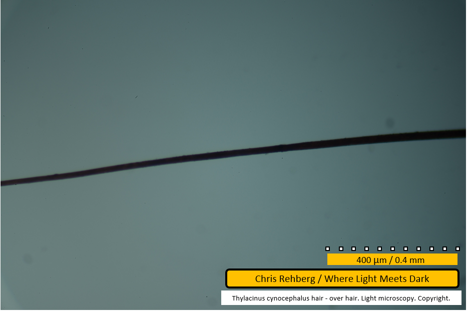
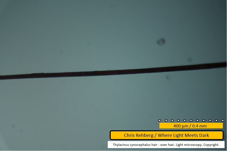
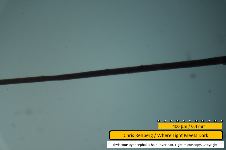
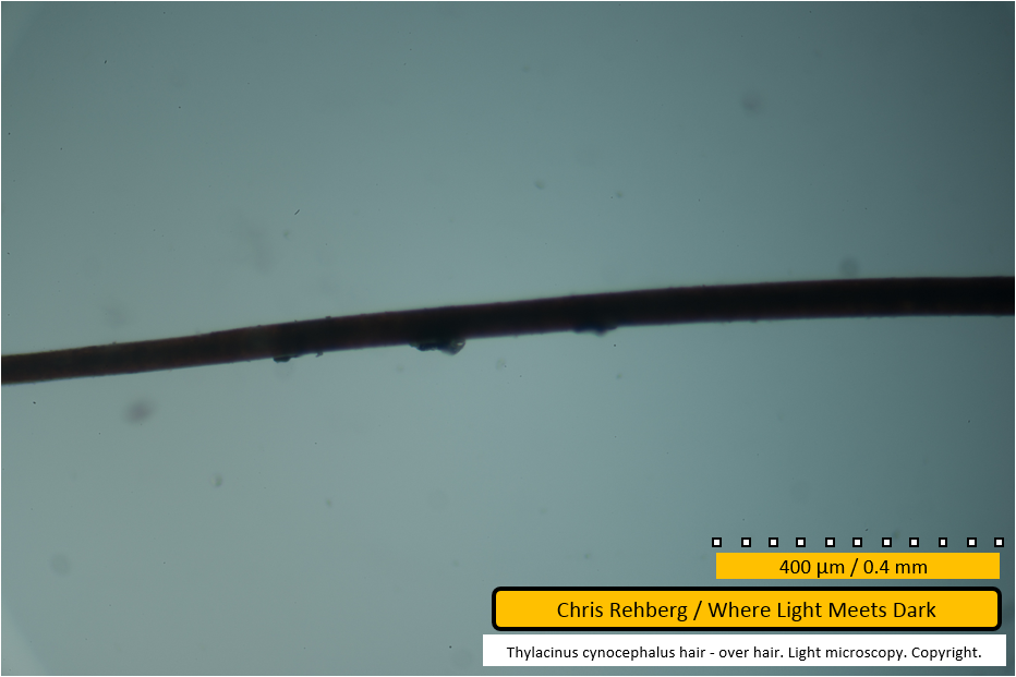
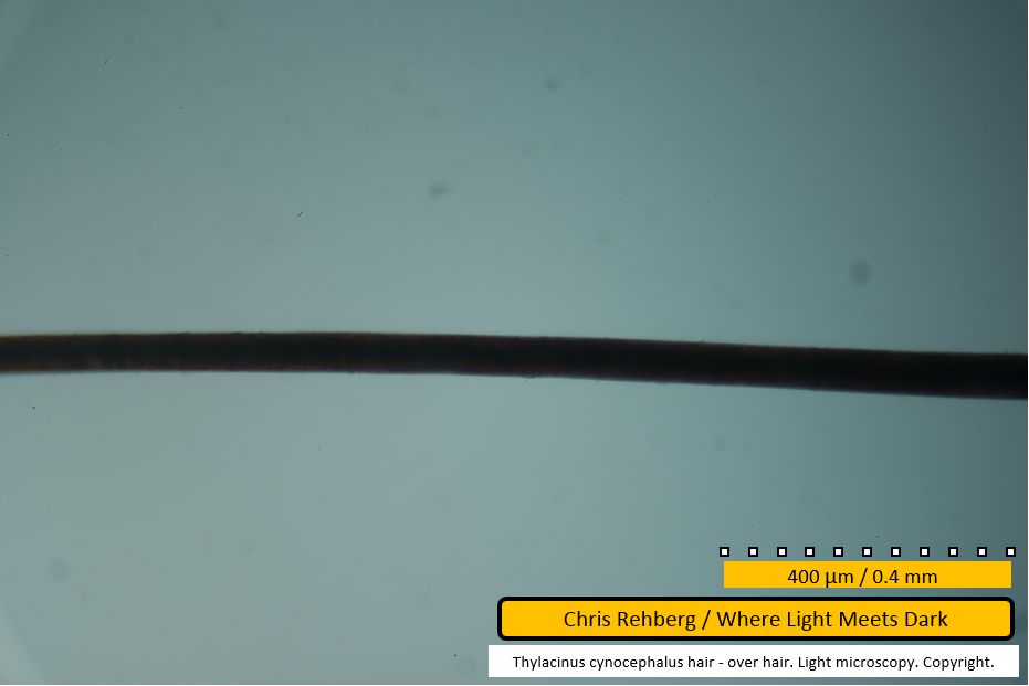
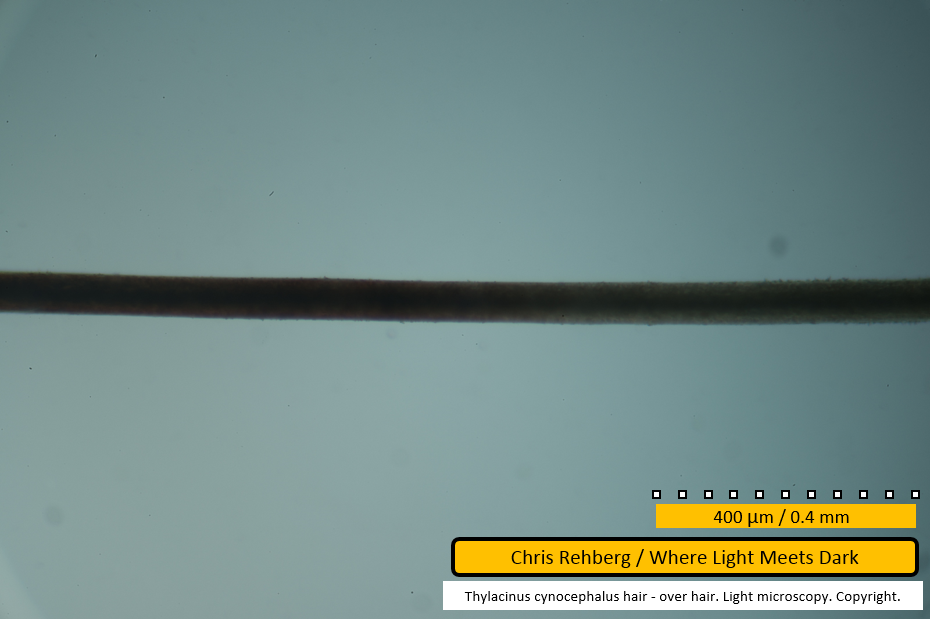
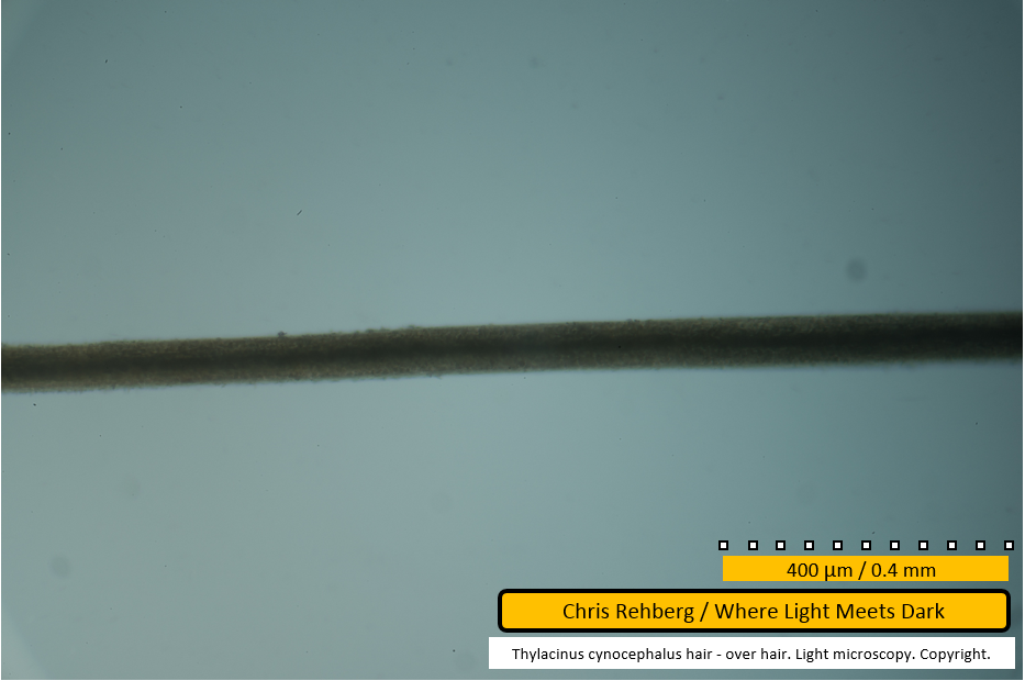
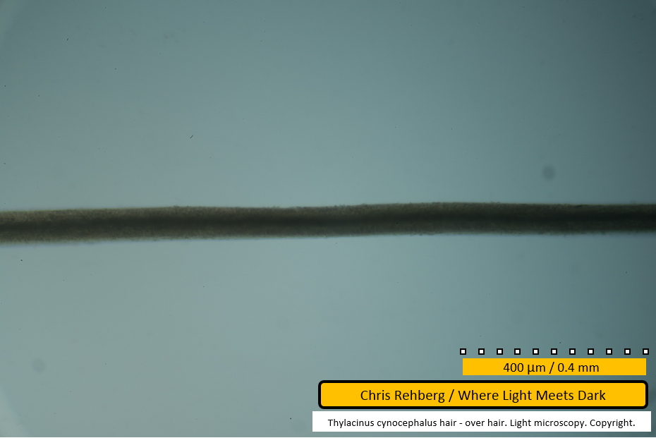
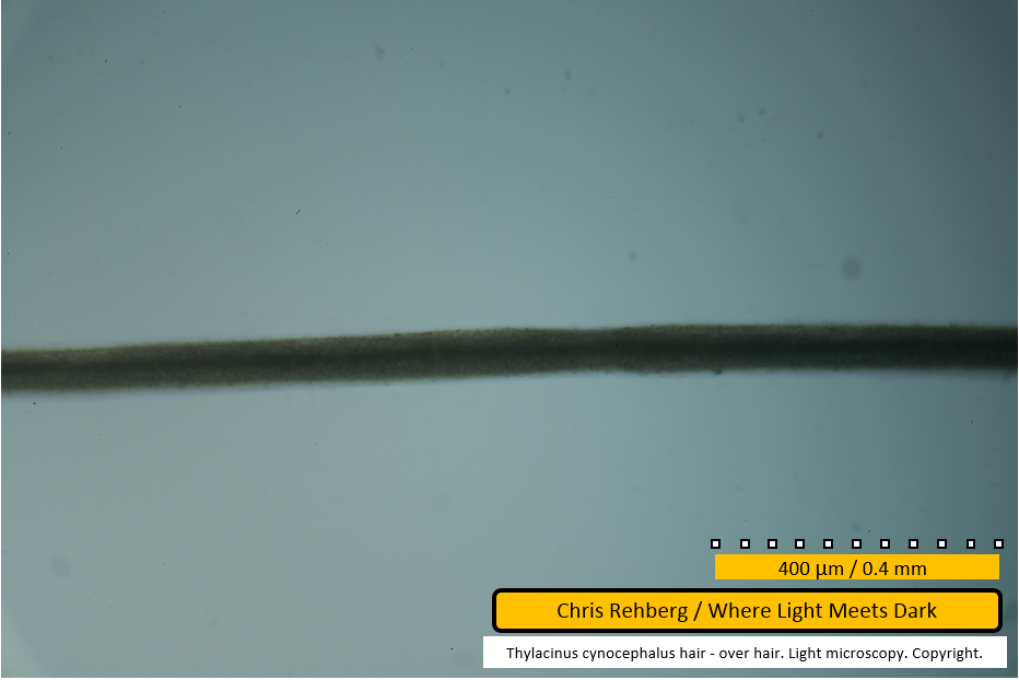
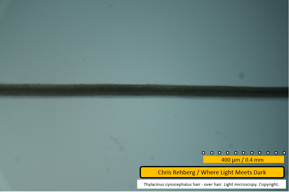
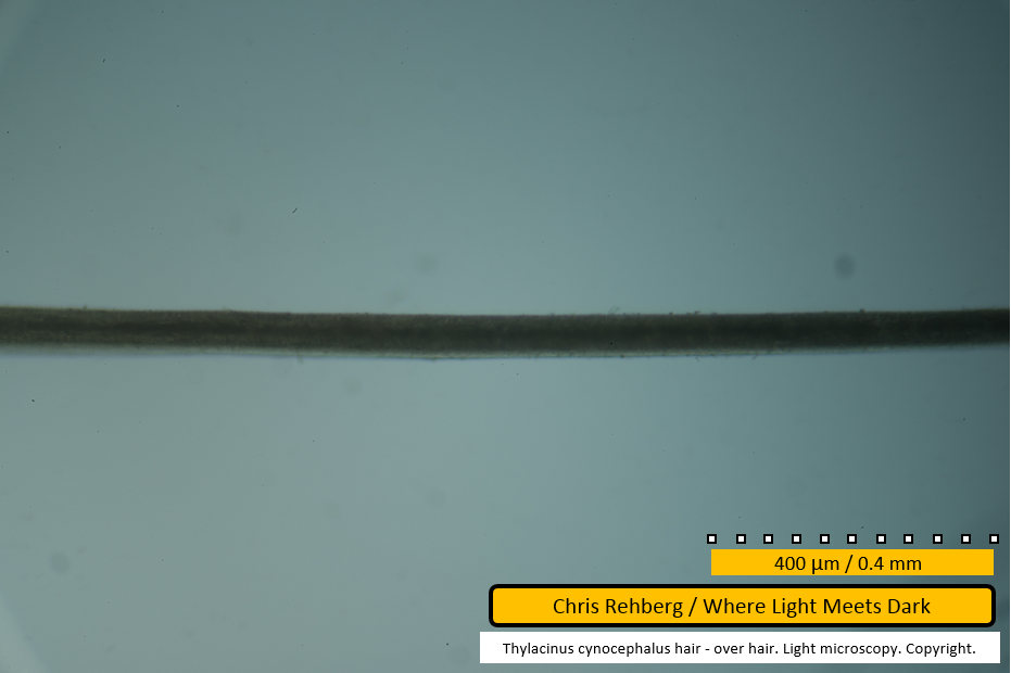
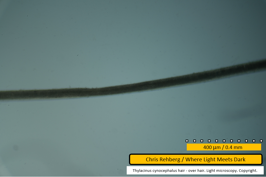
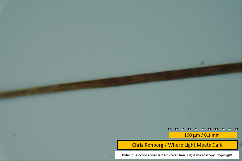
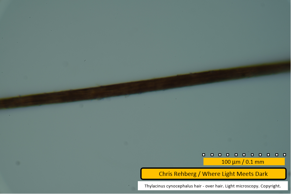
OLM Optical Light micrographs of Tasmanian tiger over hair
This is one page of results for the article OLM Optical Light micrographs of Tasmanian tiger hair.
To cite this page of results or any image within this page, see "Citing this article" at OLM Optical Light micrographs of Tasmanian tiger hair.
This page contains 37 images totaling 20MB.
Notes
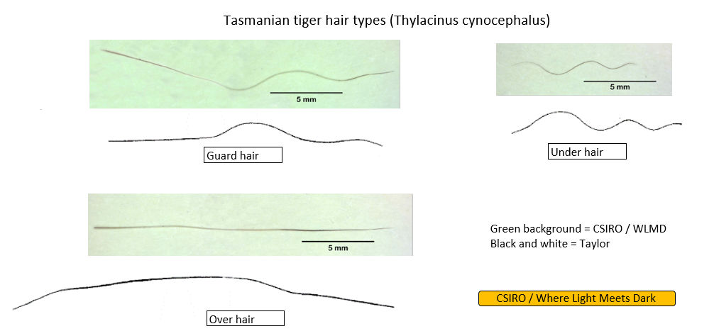
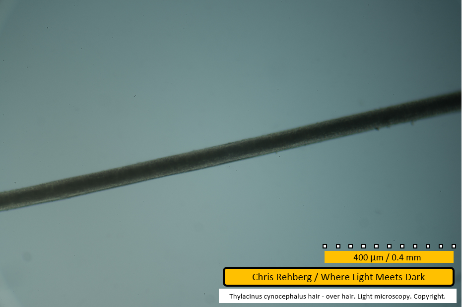
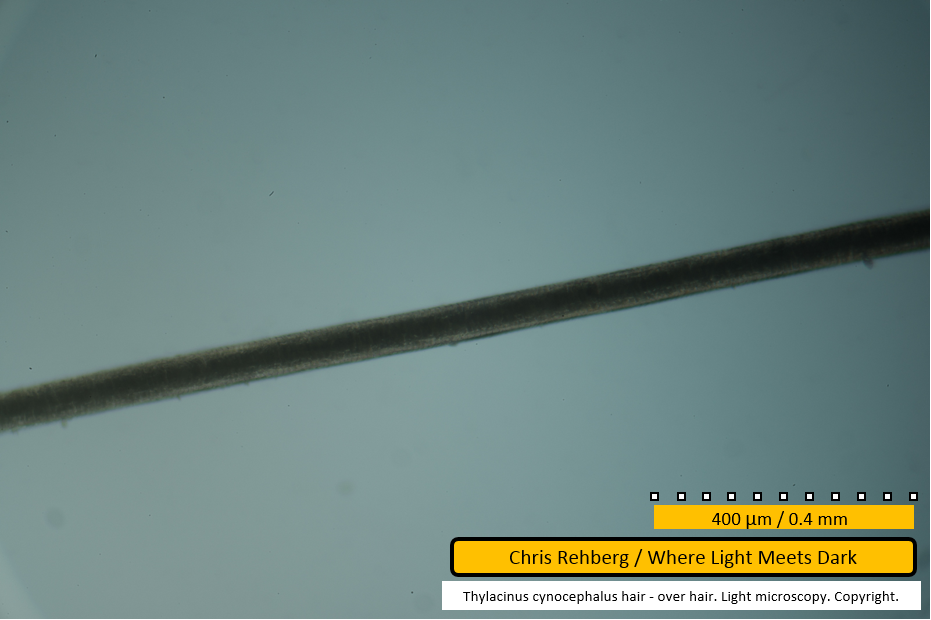
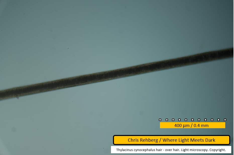
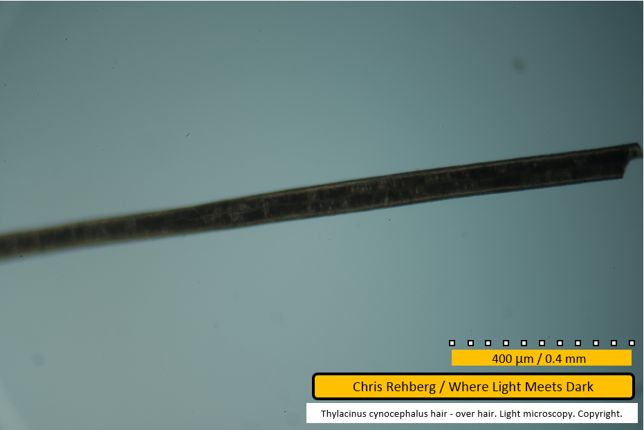
On first examining the thylacine hair sample during the SEM work, it was immediately obvious that the three thylacine hair types were present. Images of the whole fibres were presented in the results of that work and are reproduced below. Although examination of the over hair fibre showed that it was broken toward the root end, it is clear from its length - and lack of waviness throughout that length - that this was an over hair and not a guard hair.
At the time of the light microscopy work, no further over hair fibres could be found in the sample, as those fibres imaged by CSIRO had remained at the CSIRO laboratories. The reason so many guard hair fibres were examined in this study was in search of what may be an over hair fibre with a break, making it shorter. However no further over hair fibres were found. CSIRO was contacted and one of the two over hair fibres was shipped back to the author for the light microscopy work. As such, the over hair sample used in the light microscopy work is in fact the same fibre for which SEM (with and without metal coating) was carried out. As such, a direct comparison between the SEM scans and light microscopy for the exact same fibre is available, although with the fibre orientation differing. The break in the fibre is visible below, using light microscopy, from file 4040 onward and may be compared with file aa in the initial SEM results.
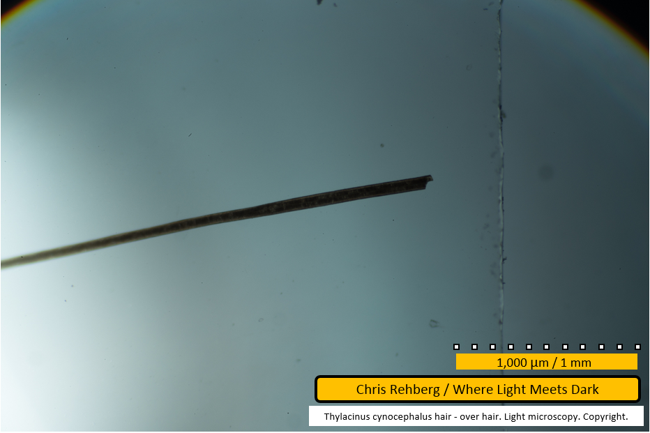
File 4013 - proximal end
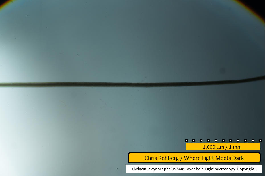
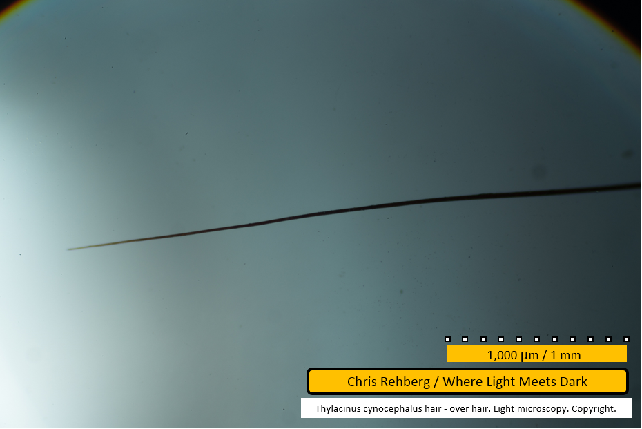
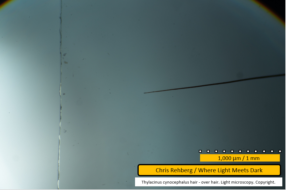
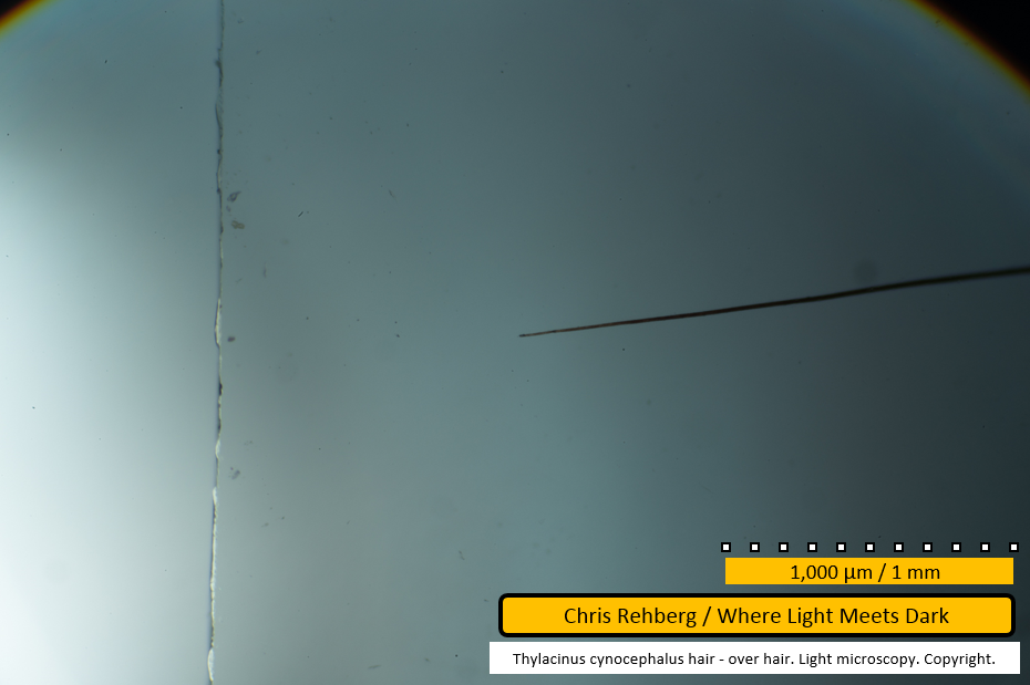
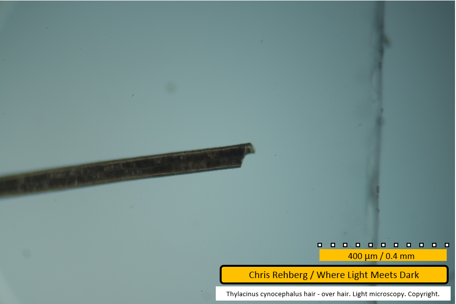
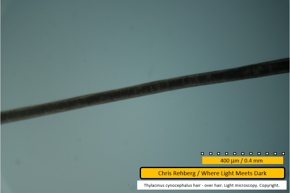
The break toward the proximal root end of the over hair fibre, as seen under lowest magnification. This, and other images showing the 1,000um/1mm scale bar were captured using 40x magnification. At this low resolution, a clearly defined medulla is present, having a somewhat patchwork appearance. This will be explored further, below. The left end of the fibre appears thinner but is out of focus. The vertical line to the right of the fibre is the edge of the cover slip on the slide. The following images through to file 4022 present the entire length of this (broken) fibre, captured at 40x magnification.
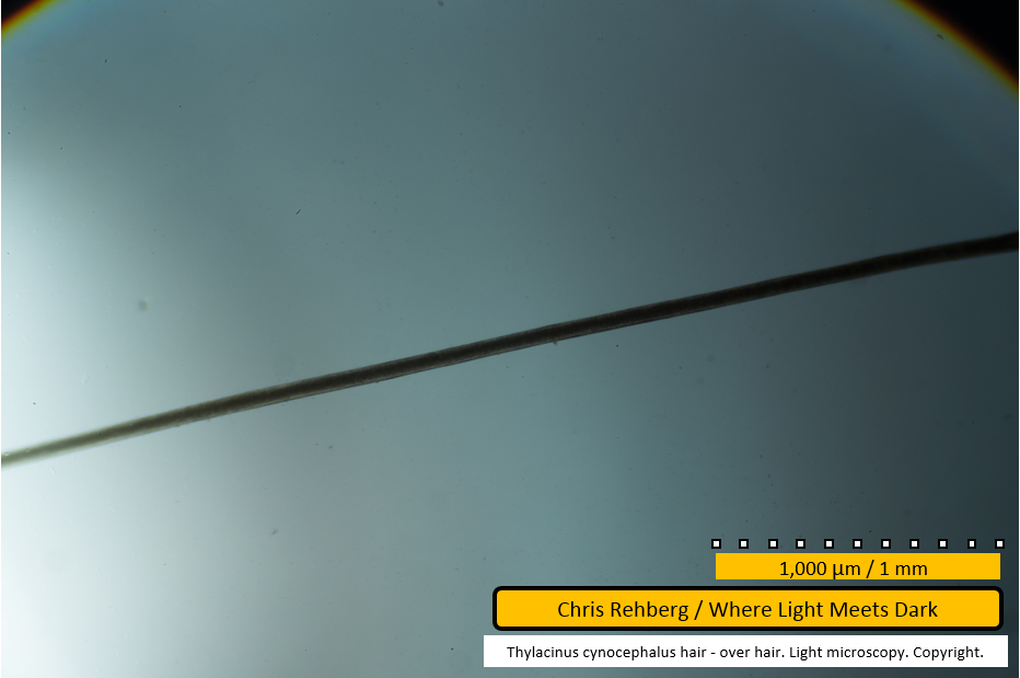
File 4014 - mid section
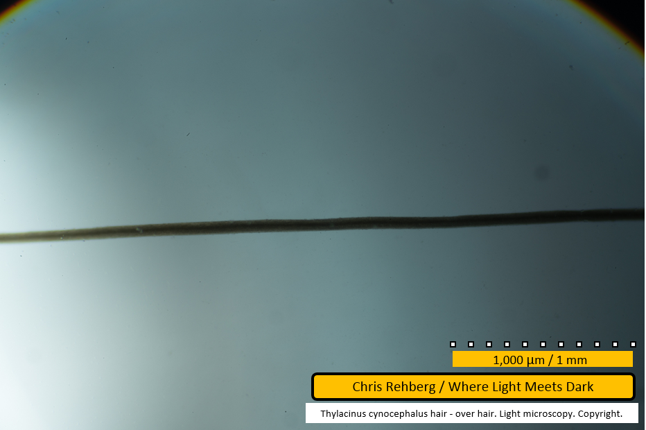
Panning toward the left, we continue to view the over hair captured at 40x magnification.
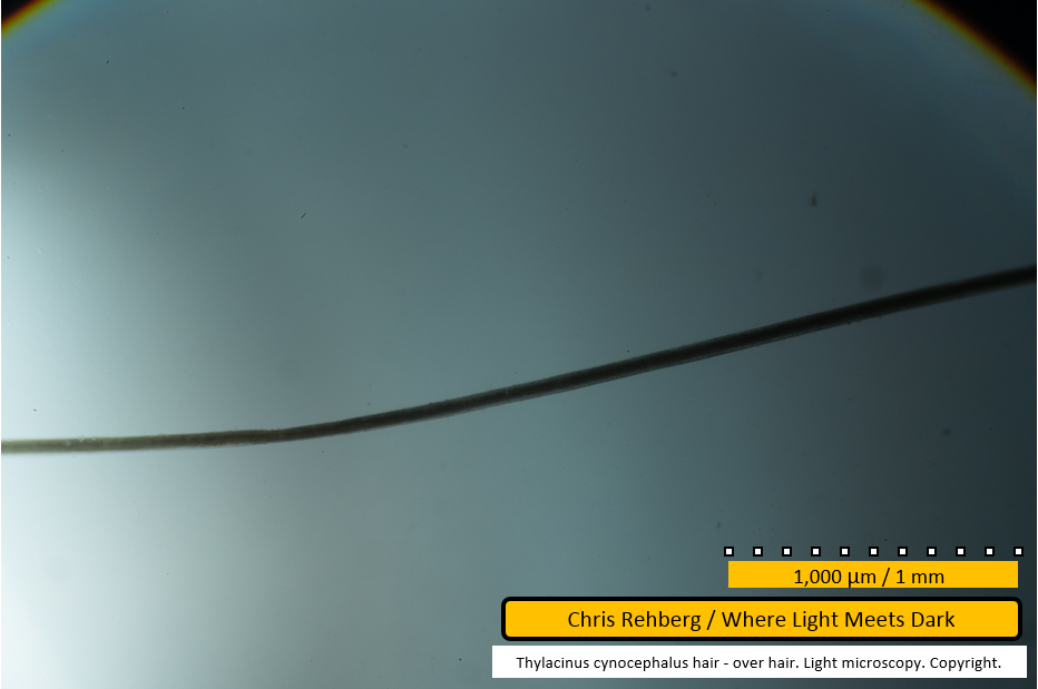
File 4015 - mid section
A slight bend in the fibre is seen as we continue panning left.
File 4016 - mid section
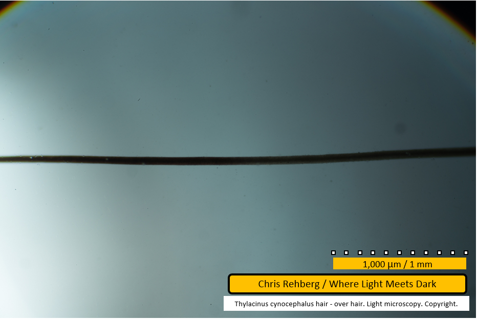
Overall the over hair fibre is quite straight, in comparison with the other two hair types for the thylacine.
File 4017 - mid section
We continue this overview of the over hair fibre, panning left.
File 4018 - mid section
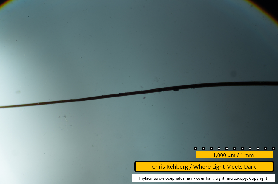
Even at this low magnification, a relative darkening of the fibre at left of frame, toward the distal end, becomes apparent.
File 4019 - mid section
In this frame the hair is clearly seen tapering toward the left. Its colouration is again darker than the earlier frames as we near the distal end.
File 4020 - distal end
The very thin distal end of the over hair, captured at lowest magnification.
File 4021 - distal end
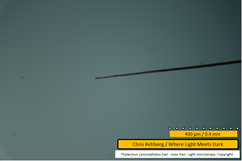
This frame presents the distal end at centre of frame where it is in better focus than in the prior image (file 4020).
File 4022 - distal end
This image is near identical to file 4021.
File 4023 - distal end
This image of the distal end was captured at a higher magnification of 100x. More detail is visible, although there is no medulla present. Some darker banding is slightly discernible in the right half of frame but does not appear to extend all the way to the end of the fibre, in common with one guard hair (file 2986, suggesting all other guard hairs terminated prior to their natural ends).
File 4024 - mid section
As we begin to traverse the fibre, panning from the distal end at left, to the broken root end, toward the right, few features are discernible in this image, captured at 100x magnification. There is the suggestion of the dark banding seen earlier on the guard hairs, in the left half of frame.
File 4025 - mid section
We continue panning right along the fibre. There is no obvious medulla in this image but the right hand side of frame is out of focus.
File 4026 - mid section
In this image, near the left edge of frame the medulla is visible as a series of small cells spanning less than 50% of the width of the fibre. Also in the left half of frame the fibre appears compressed - possibly due to manipulation with tweezers.
File 4027 - mid section
There is a suggestion of the medulla at left of frame. Dirt is adhered to the fibre, particularly along the lower edge.
File 4028 - mid section
The medulla is visible in this image as a darker central section along the fibre, but it is difficult to discern. This portion of the fibre is coloured more darkly than the remainder (in the images below) and correlates with the darker brown hair end seen in the guard hairs also.
File 4029 - mid section
This image clearly shows the more chocolate brown colour toward the over hair tip (at left) compared with the sandy or straw colour found along most of the fibre length (at right). The medulla is visible, though not sharply in focus, as continuous throughout both colour regions.
File 4030 - mid section
As we continue panning right, the paler colouration remains, providing better contrast with which to discern the medulla. At this magnification few details are visible except that the width of the medulla is proportionally less than the width seen in the guard hairs.
File 4031 - mid section
The fibre continues and remains straight.
File 4032 - mid section
The same fibre, continuing to pan to the right. The paler lines, visible against the cortext above and below the medulla in left of frame are suggestive of the scale pattern. The scale pattern will be much more clearly visible in the higher magnification images (below).
File 4033 - mid section
Again the area above and below the medulla is suggestive of the scale pattern. The medulla remains continuous and the fibre remains straight. There is a suggestion of the thinning of the medulla at right of frame.
File 4034 - mid section
The fibre continues. The medulla again shows a suggestion of thinning and possible breaks.
File 4035 - mid section
There is a slight bend in the fibre here. There is a suggestion the medulla has uniserial ladder construction in the left quarter of frame. In the right half of frame the medulla appears to occupy a greater proportion of the width of the fibre.
File 4036 - mid section
The fibre continues, showing similar appearance.
File 4038 - mid section
The fibre continues. The imbricate scale pattern is visible as faint lines on the cortex either side of the medulla.
File 4039 - mid section
The fibre continues, with broad medulla. The scale pattern is visible at far left, on the cortex and extending over the medulla. The broader pale lines traversing the medulla are suggestive of spacing between medullary cells.
File 4040 - proximal end
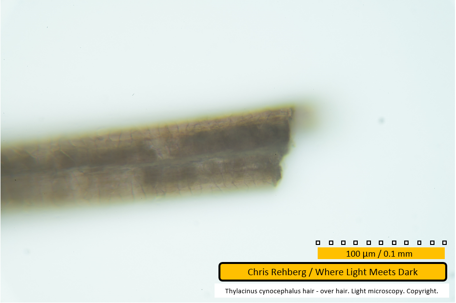
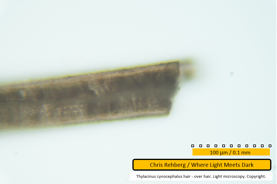
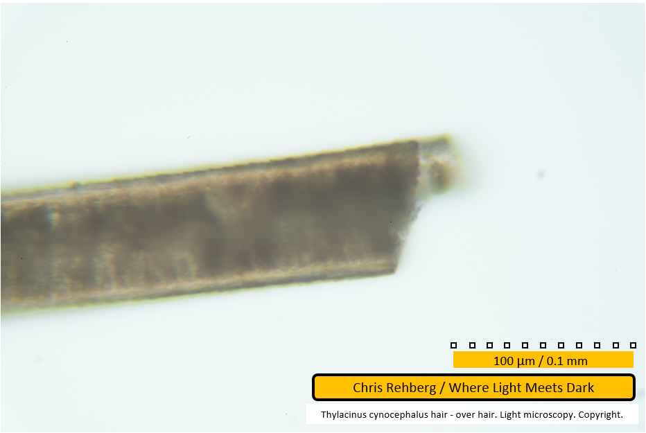
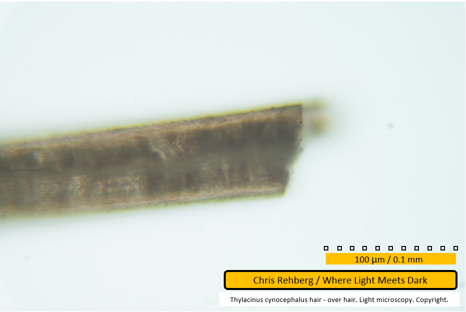
This image shows the proximal end of the fibre, although a significant break in the fibre is present. It is unknown how near to the root this break might have been. The medulla's appearance is unique here, with respect to elsewhere on this fibre or anywhere on any of the other fibres. It shows two distinct features: a line running relatively straight down the centre of the fibre, with some bends and an apparent split at far left (just before the out-of-focus region); and paler regions in the medulla interpreted as vacuolation - sometimes above this line, sometimes below and sometimes both above and below. The cause of this line is unknown. The vacuolation gives the appearance of there being two rows of cells - one above, and one below the line - but it would seem unusual that the hair should be coincidentally oriented to show these two rows of cells exactly one above the other (and not one in front of the other, or some orientation in between). Given the proximity of these features to the break in the hair there may be an association between them: the features may be a result of detrioration of the hair following the break, or the features may suggest an internal deterioration leading to weakness of the fibre at this point, resulting in the break. An examination of the broken end under higher magnification (files 4042 - 4045) suggests the line is at the surface of the fibre, as opposed to central. It seems probable the line represents a splitting of the fibre, likely as a result of the same pressures leading to the break, but these considerations are speculative.
File 4041 - proximal end
This image shows the break at centre of frame.
File 4042 - proximal end
This image of the broken proximal end of the over hair was captured at 400x magnification. The scale pattern is visible against the cortex above and below the darker medulla which runs along the centre of the fibre. This and the following three images (files 4043 - 4045) show the same point with differing focal depths in order to highlight different features. Note in this image that the scale pattern cannot be seen at the extreme edge of the fibre (which is out of focus). The central line discussed under file 4040 does appear to be in focus, suggesting this feature is at the hair surface. (Compare with file 4044, where the scale pattern at the extreme edge is in focus and the central line is out of focus).
A direct comparison with the scanning electron micrographs of the earlier research is not possible as the SEM work imaged the opposite side of the fibre. This is apparent given that in this image the protrusion at the break is out of focus, suggesting it is at the bottom of the fibre, looking down onto the fibre, as here. File aa in the SEM work clearly shows this protrusion uppermost as the scanner looks down on the fibre.
File 4043 - proximal end
The same broken end with a different focal depth. In comparison with the prior image, the scale pattern near the very edge of the fibre begins to take on a spinuous appearance. It is in fact imbricate but one scale does overlap the next, leading to the pattern seen here. As with earlier images, with the scales apparently raising toward the left of frame, it can be concluded the distal end is toward left. Note that the central line is less in focus than in the prior image (file 40420).
File 4044 - proximal end
In this image the scales at the edge of the fibre are in focus and the manner in which one scale overlaps the next is apparent (see also notes on file 4043). With focus sharply on these edge scales, the focal depth is at the centre of the fibre (looking down onto the fibre). The central line discussed earlier is a blur in this image, and out of focus, again reinforcing the conclusion that it exists on the surface of the fibre facing the camera, as opposed to existing at the centre of the fibre (despite it running centrally along the length of the fibre in this orientation). Some of the vacuolation (see notes on file 4040) is visible here, both above and below the (blurred) centre line.
File 4045 - proximal end
The broken end with another variation to the focal depth.
File 4046 - mid section
This image shows a portion of the mid section, captured at the lower magnification of 100x. The central line is visible at far right of frame. The vacuolation is visible in association with the central line but also further left (near centre of frame) where the line is not apparent.
File 4047 - distal end
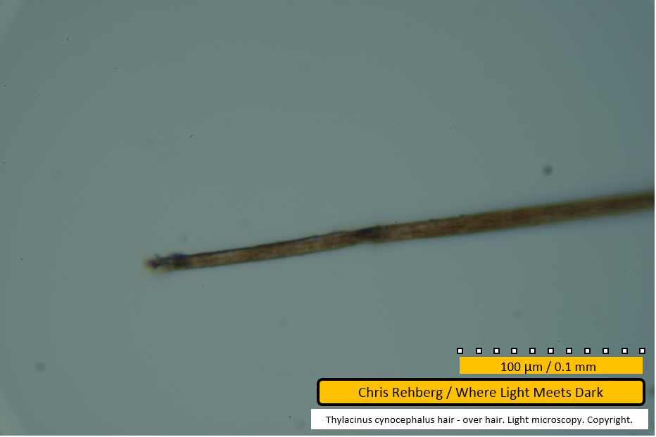
This and the remaining images were captured at 400x magnification and show the distal end of the over hair. There is a dark, near black mark at the very tip, similar to what was seen in one of the guard hairs. In this case there is a second greyish-black mark further along the fibre. There is no medulla at this point in the fibre. The general colouration is sandy brown.
File 4048 - mid section
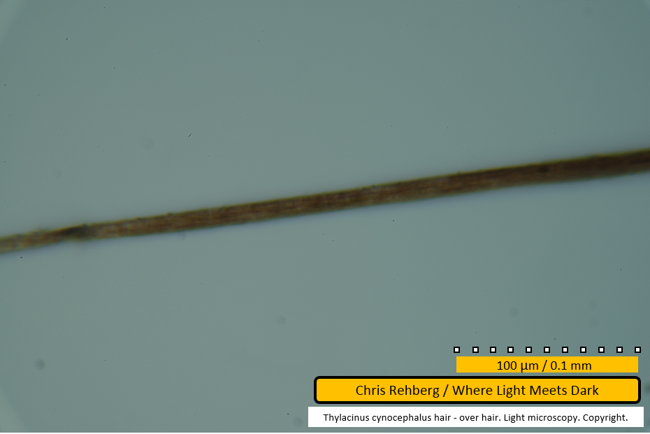
As we pan to the right the tip of the over hair remains sandy brown in colouration, without a medulla.
File 4049 - mid section
As we continue to pan right the fibre becomes a darker brown. Broad dark brown stripes are apparent traversing the fibre at far right, in common with the guard hairs. In four of the five guard hairs, this banding pattern extended almost to the terminus of the fibres but in one guard hair an extended, unbanded, straw coloured tip was observed (file 2986) suggesting that the tip of over and guard hairs is similar in appearance (when not broken). As noted earlier, the distal end of guard hairs is wavy (immediately back from the tip), whereas in the over hair, as seen in these images, it is straight.
File 4050 - mid section
The banding pattern continues along this dark brown portion near the distal end of the over hair fibre.
Under hair results
Go to Under Hair Results
Guard hair results
Go to Guard Hair Results
Main article
Go to Main Article
