OLM Optical light micrographs of Tasmanian tiger under hair
This is one page of results for the article OLM Optical Light micrographs of Tasmanian tiger hair.
To cite this page of results or any image within this page, see "Citing this article" at OLM Optical Light micrographs of Tasmanian tiger hair.
This page contains 28 images totaling 16MB.
File 2948 - proximal end
This photo shows the proximal end of an under hair. The spinuous scales are clearly visible in left half of frame and point toward left of screen, indicating the distal end of the fibre is toward the left. In this photo the medulla is absent at the root, broken near centre of frame, then continuous and forms a unicellular ladder in the left portion of the frame.
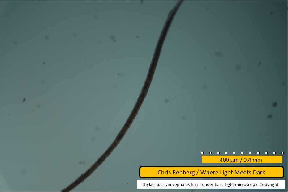
This, and subsequent images showing the 400um scale bar, were captured at approximately 100x enlargement.
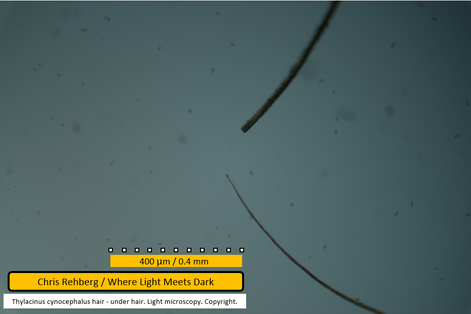
File 2949 - two under hairs - distal ends
Files 2948 and 2959 illustrate the proximal ends of the two under hair fibres, neither of which is seen in this image. As such, this image illustrates the distal ends of both under hair fibres. The uppermost fibre has been cut and its long, tapering tip has been removed, accounting for the discrepancy in diameter at these ends. In the uppermost fibre some medullary cells are visible.
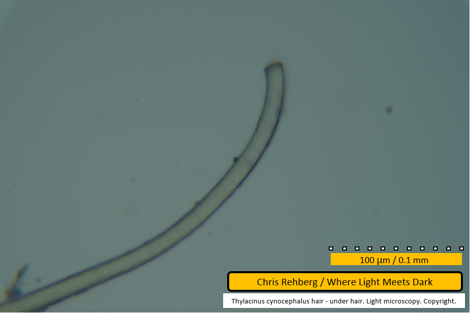
Little detail is shown in this shot but the comparative difference in diameter between the two fibres at their ends is obvious.
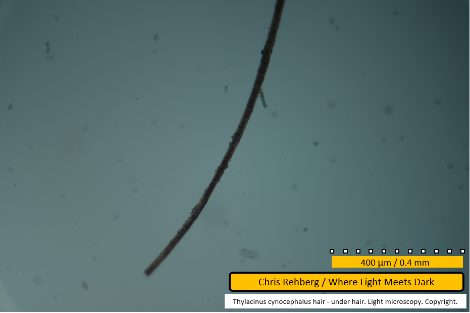
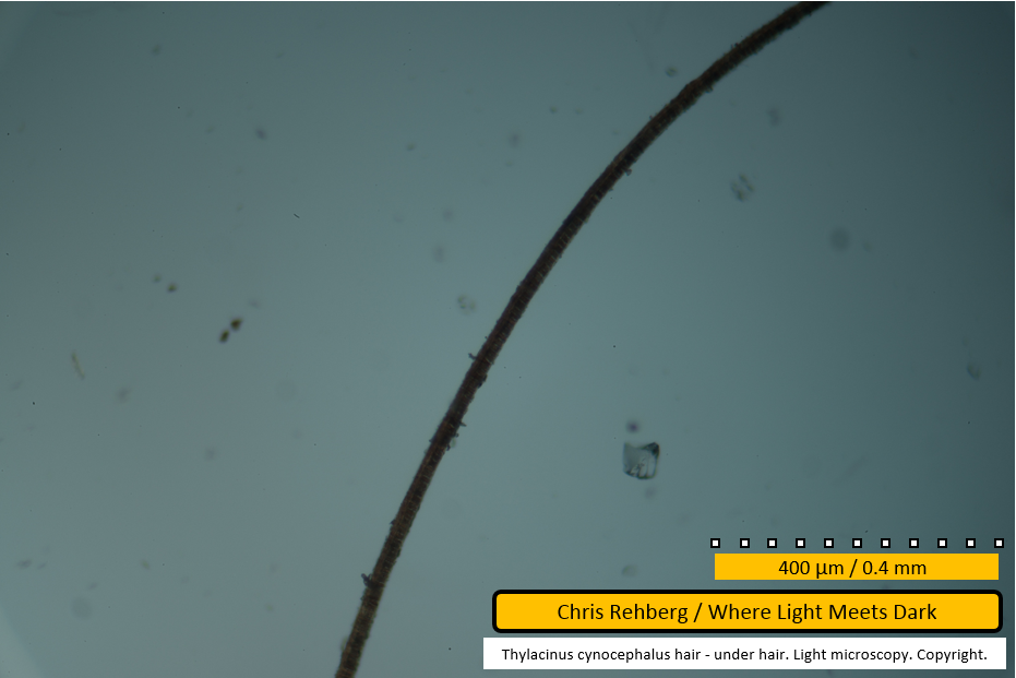
File 2950 - distal end
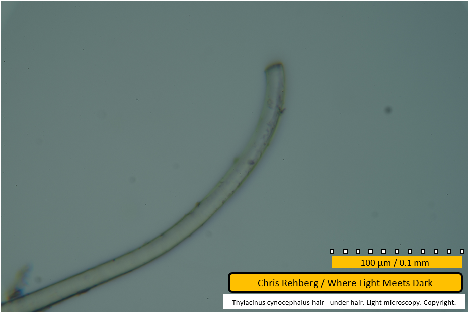
The medullary cell pattern becomes obvious in this image; the medulla consists of small "blocks" running down the centre of the fibre forming a unicellular ladder, shown here as being darker than the edges of the fibre. Toward the end of the fibre the lattice is less regular (ie. broken).
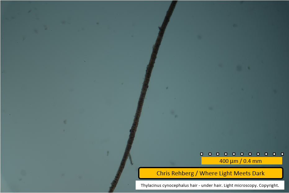
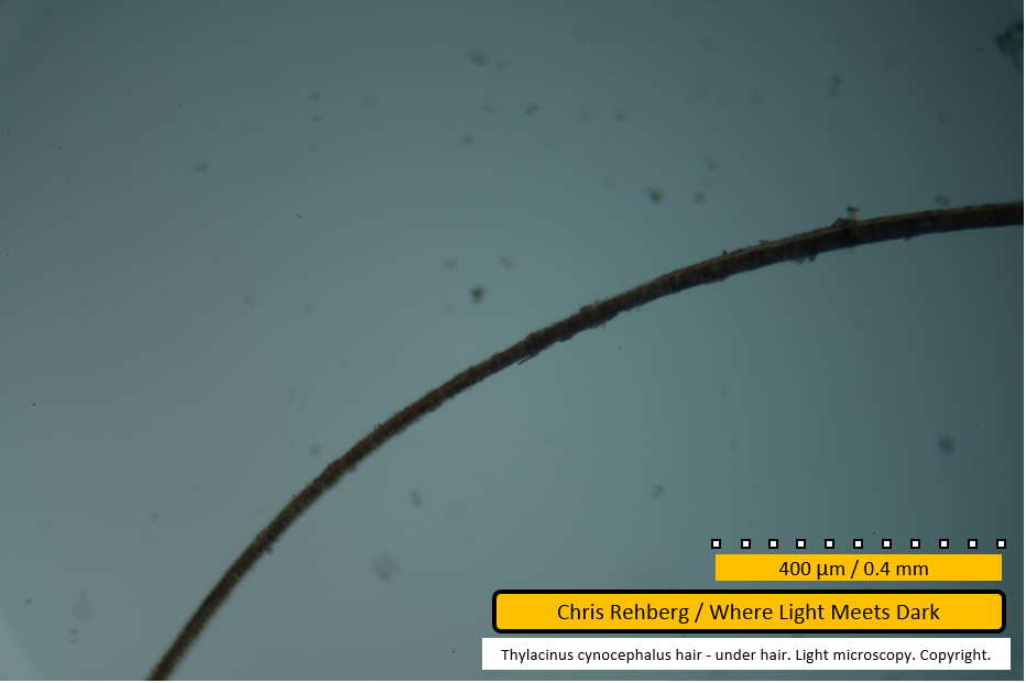
File 2951 - mid section
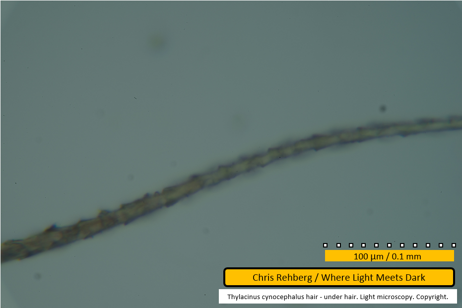
Note the protrusion attached to the fibre near the bottom of the image - this is also visible in the previous image (file 2950). The medulla is again clearly seen as a series of blocks along the centre of the fibre. Dirt particles are visible adhered to the fibre at its edges.
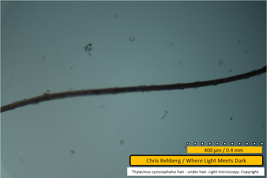
File 2952 - mid section
Further along the same fibre as shown in the previous image (file 2951).
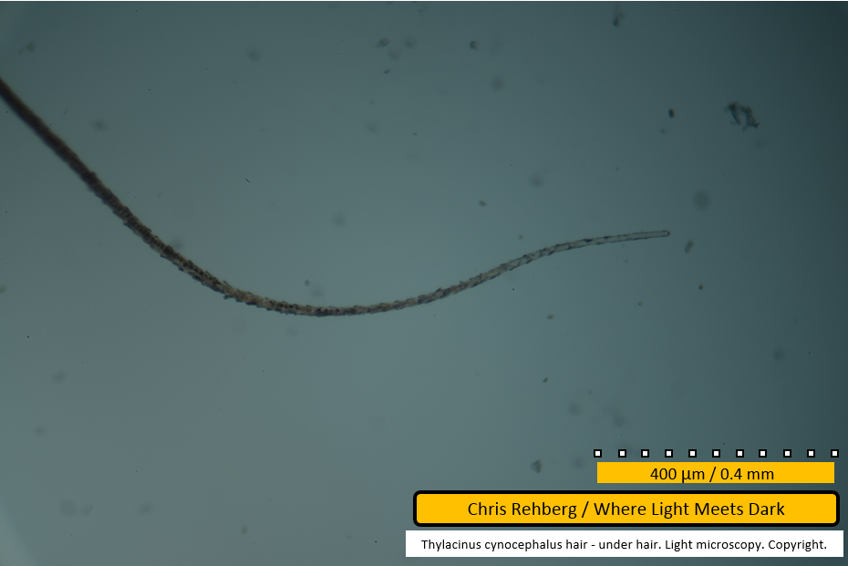
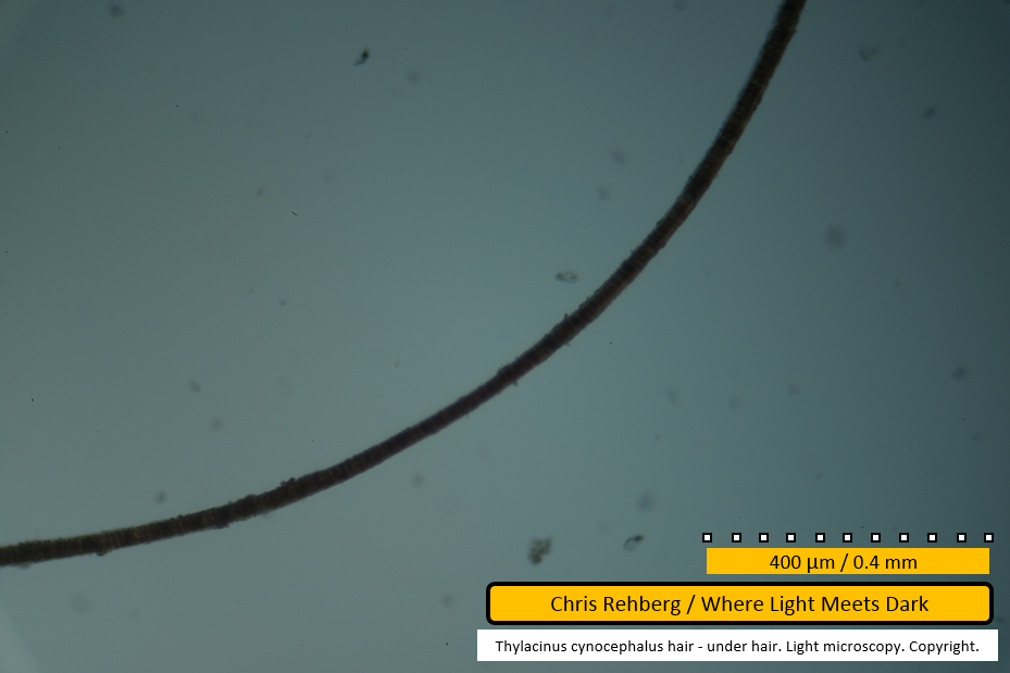
File 2953 - mid section
Further along the same fibre as that shown in the previous image (file 2952). The curve of the fibre can be discerned by tracing through the last four images (2950, 2951, 2952, 2953) as it moves from near vertical in the frame to near horizontal. The fibre diameter is notably thicker at right of frame.
File 2954 - mid section
The same fibre continues.
File 2955 - mid section
The same fibre continues to curve sinusoidally. The length of each segment in the medulla is relatively consistent despite the fibre diameter varying slightly.
File 2956 - mid section
The same fibre continues. The edge of the fibre near centre of frame is suggestive of scales but this is not definitive.
File 2957 - mid section
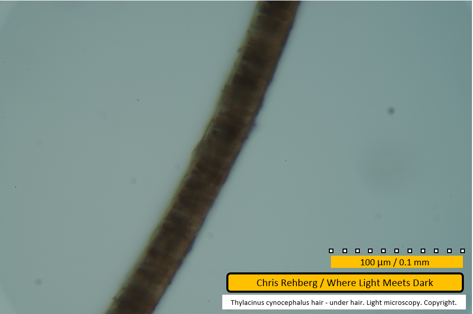
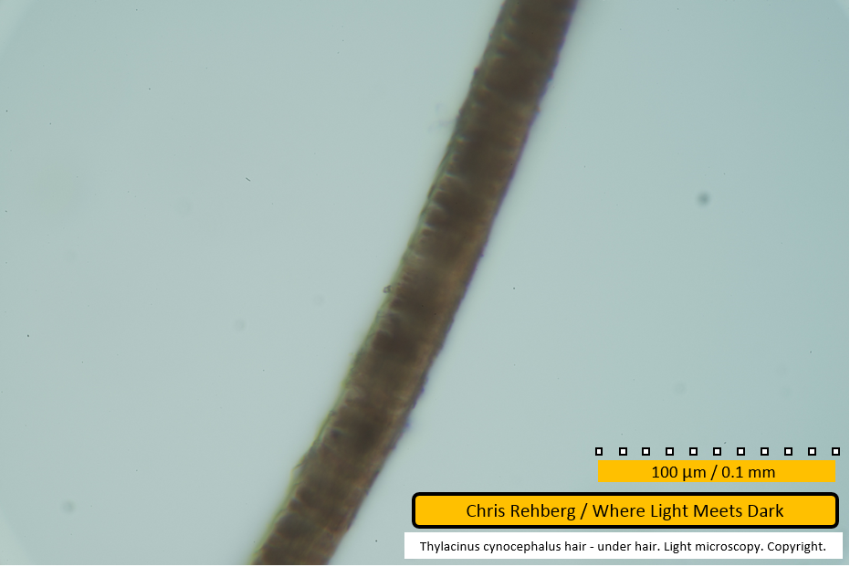
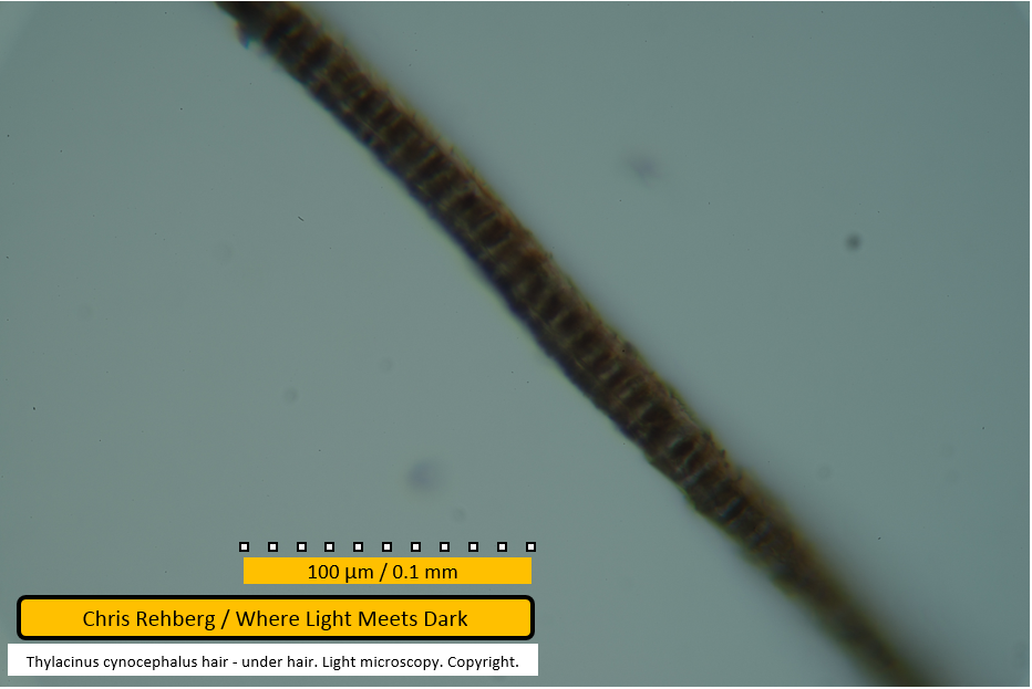
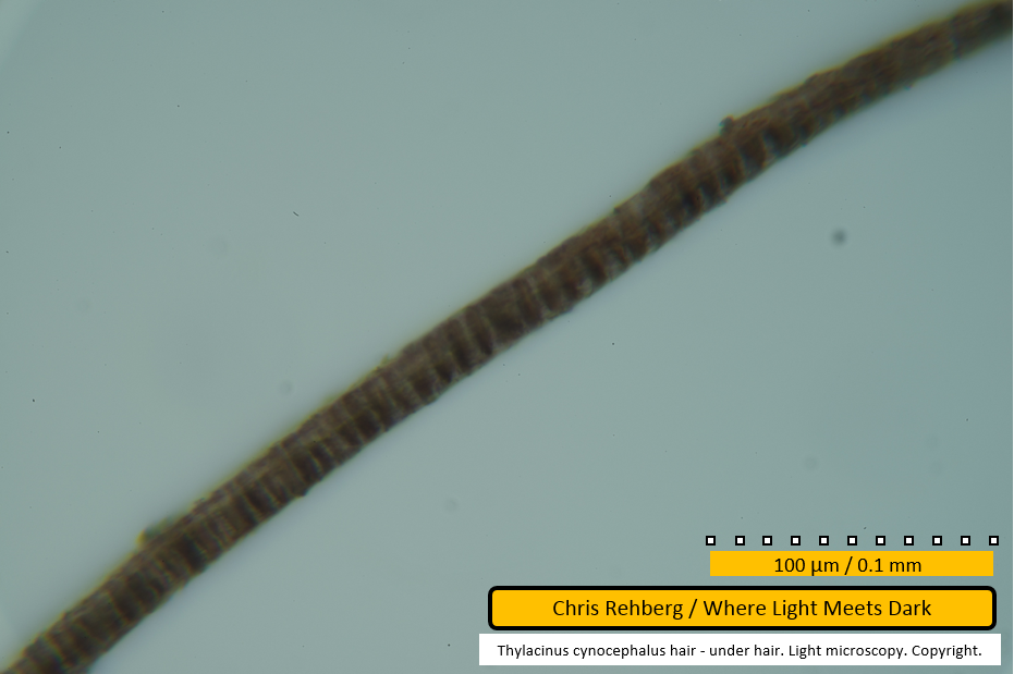
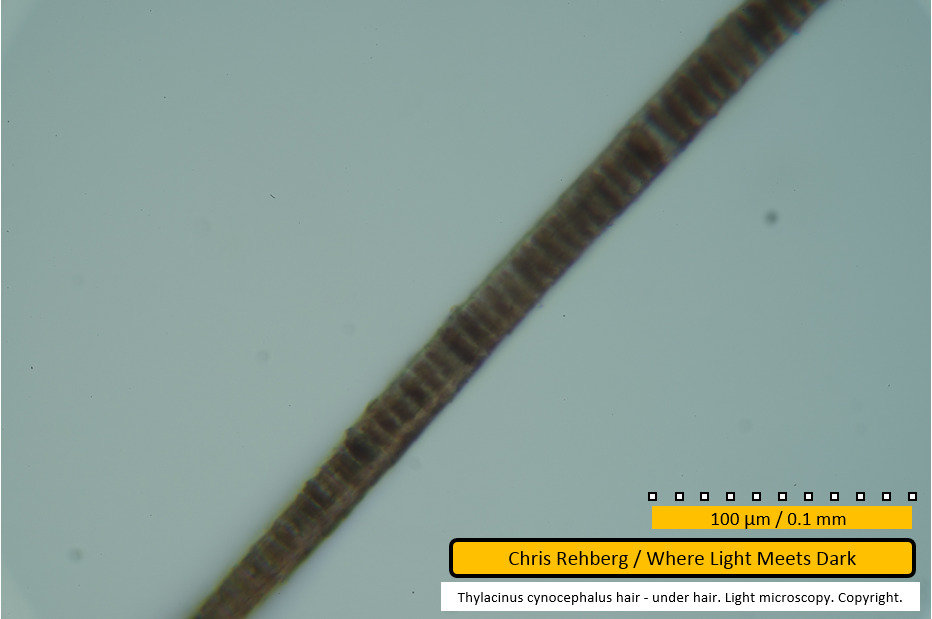
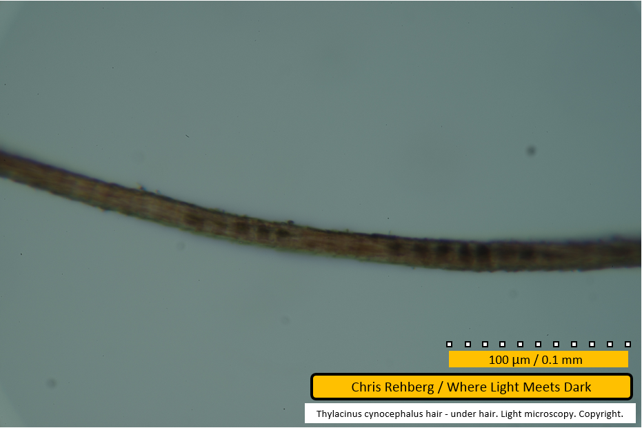
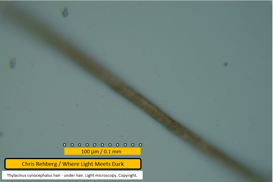
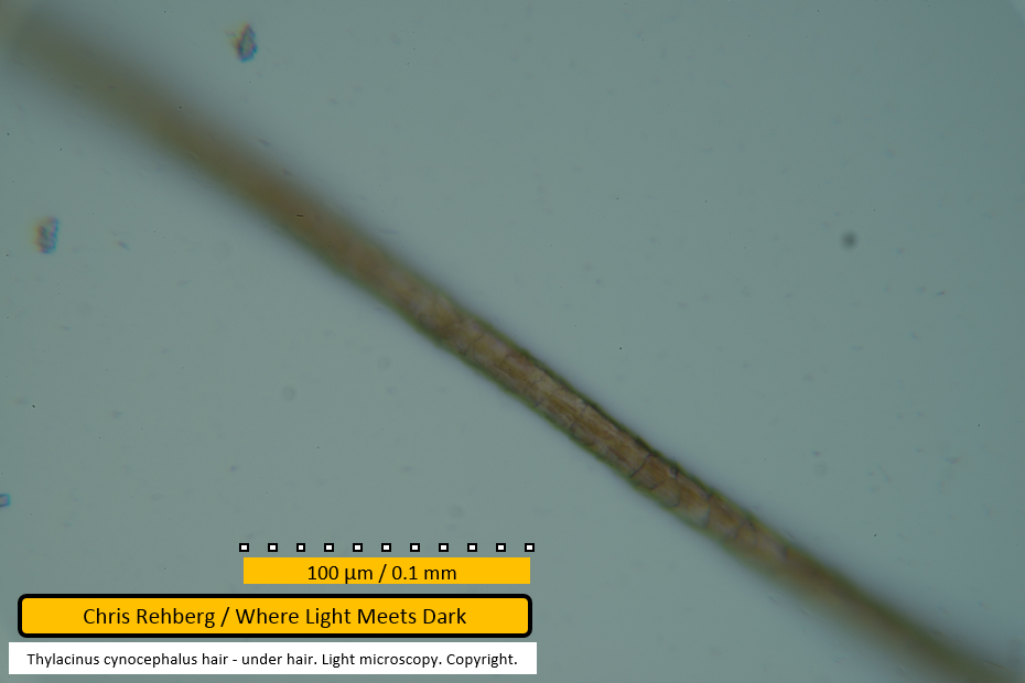
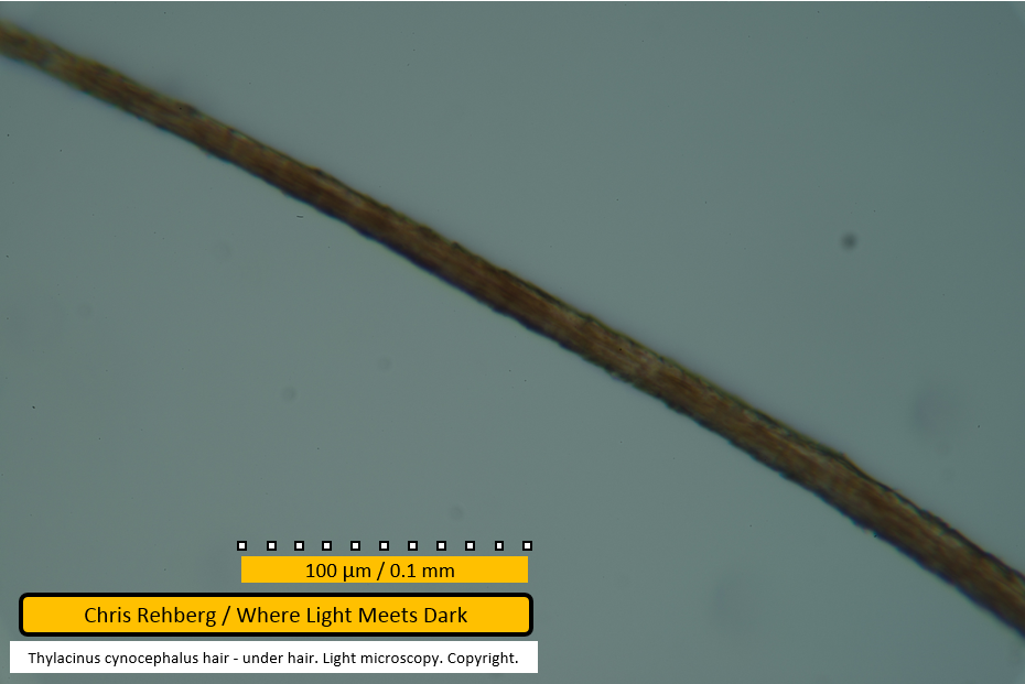
In this image the 40x objective was used and enlargement at time of capture was approx 400x. Subsequent images showing the 100um scale bar were also captured at 400x.
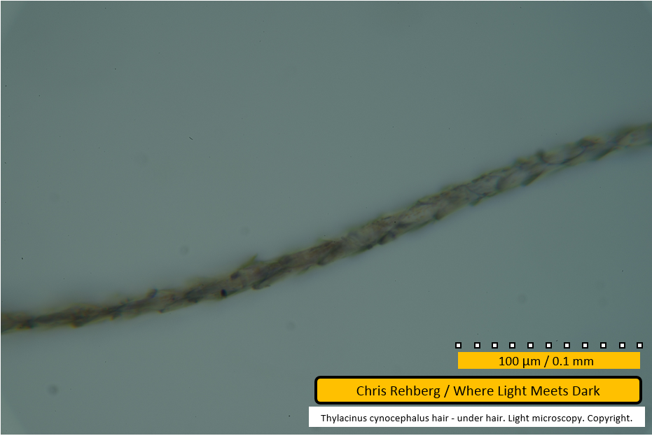
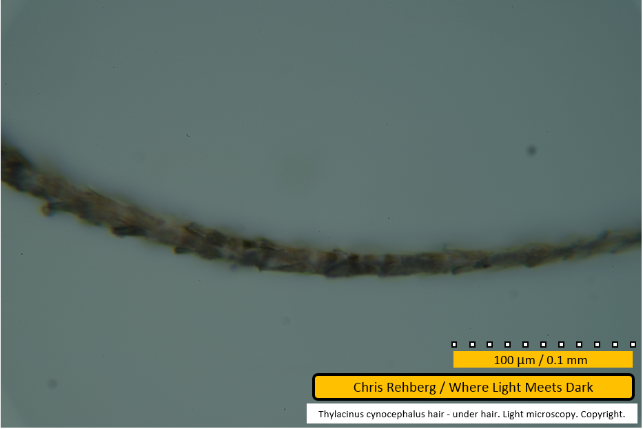
Very thin pale lines may be seen traversing the fibre - these represent scale edges on the fibre surface.
File 2959 - proximal end
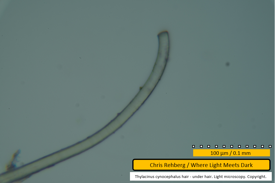
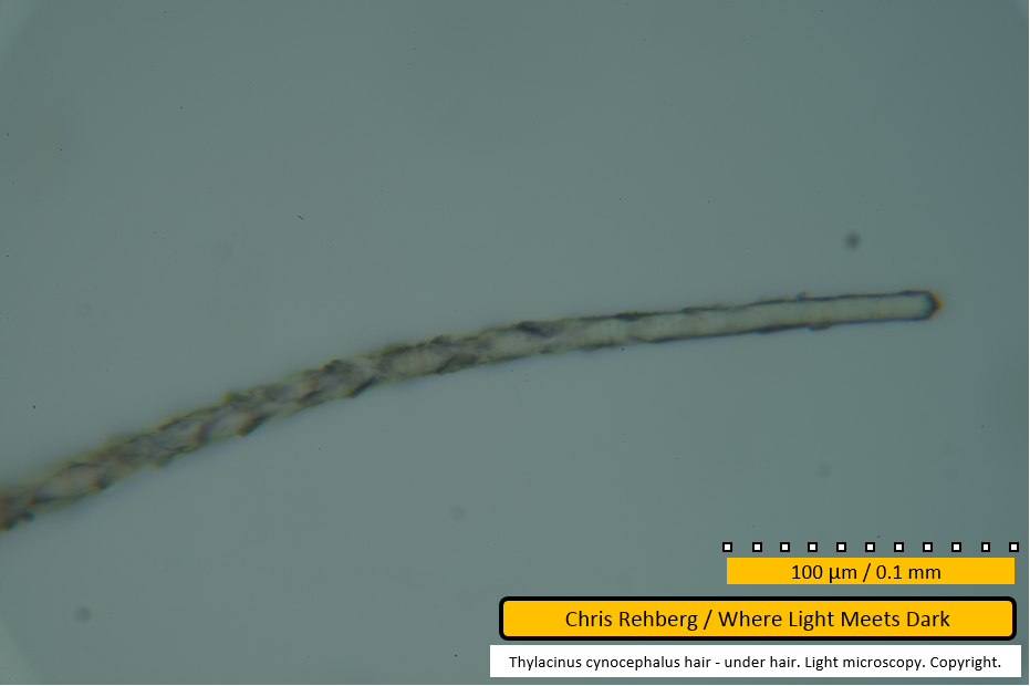
The end of the same fibre is translucent due to lack of medulla. Very faint darker lines may be seen traversing across the fibre - these represent the scale edges. The distance between scale edges is considerably less than 10um.
File 2960 - mid section
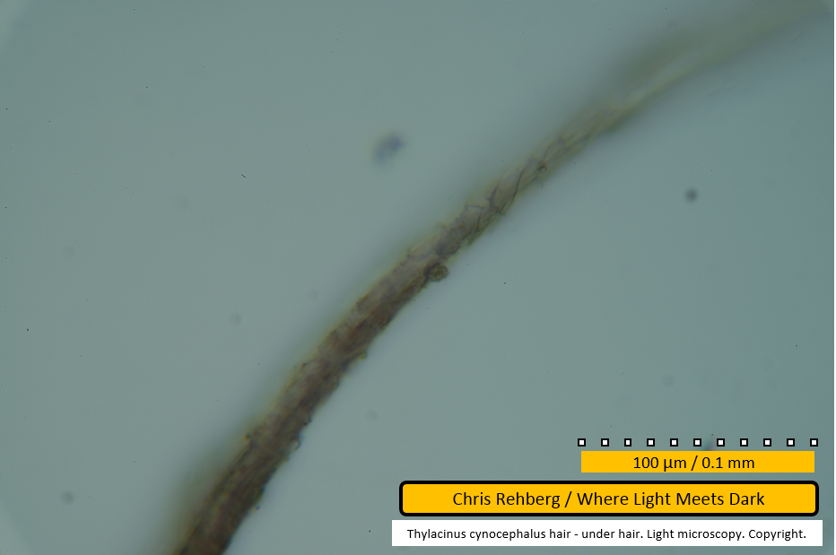
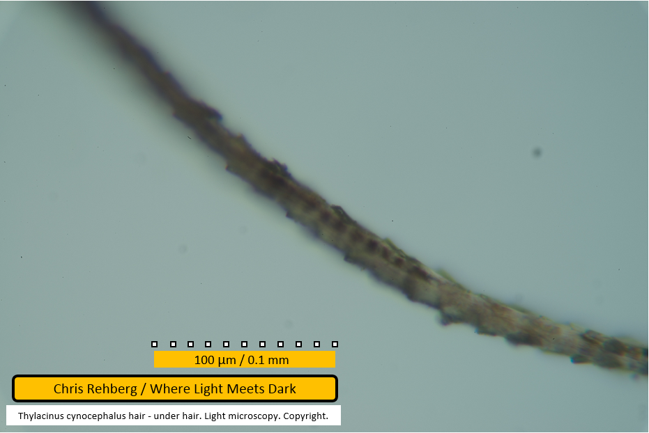
A portion of an under hair showing scale pattern (darker diagonal lines) along a short length. The surface of the fibre is sharply in focus near centre of frame, yet grossly out of focus only 50 micrometres along.
File 2961 - mid section
Another close-up showing scale pattern - more notable near edges - as paler lines traversing the fibre.
File 2963 - proximal end
The proximal end shown earlier (in file 2959) is shown again for comparison with file 2964 in which a different focal depth is used.
File 2964 - proximal end
The same fibre end as file 2963 but using a different focal depth.
File 2965 - mid section
This segment shows scales clearly lifted at the edges. As these scales point toward the left of frame, the distal end of the fibre lies toward the left and the root lies toward the right. The left edge of the image shows the fibre being closer to in-focus than at the right edge. This image demonstrates how sharply focused images are difficult at high resolution on whole-fibre slide mounts.
File 2966 - proximal end
In this image the scales can again be seen lifting off the fibre edge. They "point" toward the left, implying the distal end is toward the left and that this shows the proximal end.
File 2967 - mid section
Panning left along the same fibre, the scale pattern is still visible.
File 2968 - mid section
Continuing to pan left along the same fibre.
File 2970 - mid section
Continuing to pan left along the same fibre. Near centre of frame can be seen a number of darker segments within the fibre - these are the beginnings of the medullary core. At this point in the fibre they do not span more than one third of the fibre's diameter. Although the upper-left portion of the frame is out of focus it can be seen that the left end of the fibre in this image is clearly darker than the right end. This is due to the presence of the medulla toward the left. The next image (file 2971), taken further along the same fibre, shows the medulla spanning over 75% of the width of the fibre.
File 2971 - mid section
Panning further left, the medulla is seen here clearly spanning almost 100% of the diameter of the fibre.
File 2972 - mid section
Again the segmented medullary core is clearly apparent.
File 2973 - mid section
The uniserial ladder medulla remains clearly evident. There is a small gap between two of the cells in the medulla near top of frame.
File 2974 - mid section
The medulla is discontinuous at this point as we begin to approach the distal end of the fibre (toward left).
File 2975 - mid section
This and the next frame (file 2976) show the same portion of the fibre at different focal depths; again to illustrate that the apparent features visible within the frame depend on focal depth. The scale pattern is clearly seen as the medulla is absent.
File 2976 - mid section
This and the previous frame (file 2975) show the same portion of the fibre at different focal depths; again to illustrate that the apparent features visible within the frame depend on focal depth. The scale pattern is clearly seen as the medulla is absent.
File 2977 - mid section
The distal end of the fibre begins to taper (toward left). Where the scale pattern appears imbricate and on the fibre surface in the prior two images (files 2975 and 2976), here they are visible along the edges of the fibre - not due to any difference in the fibre's structure, but due to a focal depth that rests on the fibre's centre (here) compared with its surface (in the two prior images).
File 2978 - distal end
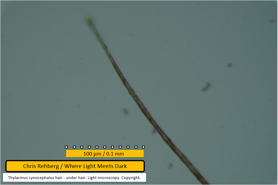
Just out of focus, it can be seen that the distal end has a frayed appearance. This is just perceptible in the lower magnification micrograph of file 2949. As you examine the fibre along its length, portions appear to show a darker brown colouration while other areas show a lighter greyish brown or, at the very tip, a straw colour. The darker brown here is not due to medullary cells; rather, it is due to pigmentation in the fibre's cortex.
Guard hair results
Go to the Guard Hair Results
Over hair results
Go to the Over Hair Results
Main article
Go to the Main Article
