Emmerichs thylacine - first comparison
Let the games begin! (part 2)
What can we do with scans of newspaper clippings of digital images? Clearly if you've seen the images at Cryptomundo (be sure to click on them for enlargements), their quality leaves a lot to be desired.
However, as I've argued in the commentary at Cryptomundo and here at Where Light Meets Dark, the composition of the images gives us plenty to work with, but only because we have two photos which have been taken moments apart.
[Side note: The analysis presented here is being conducted piece-wise, in that I will publish results as I get them. Analysing images in this manner takes time, but with the discussion running hot amongst cryptozoologists worldwide, I felt it pertinent to get the argument out there for discussion.]
Apples with Apples
The first obstacle we have to contend with is to ensure the images are resized so that they are to scale. In order to do so, we need to find an element which is present in both photos. Fortunately, there is just such an element - a leaf, which I will call L1 (leaf 1).
The smaller image at Cryptomundo will be called image S. The bigger image at Cryptomundo will be called image B. (The term "larger" was avoided to avoid confusion with using the abbreviation 'L', which in this discussion refers to a leaf).
Figure 01 - A close up of both images showing leaf L1.
A line was drawn on image B which spans leaf BL1. The image was zoomed to 200% (x2) and the line adjusted until its ends were directly over the leaf edges at relatively identifiable points. A copy of the line was positioned over image S. Image S was iteratively proportionally scaled until the line spanned the leaf SL1 in the same way as the first image. Image S was zoomed to 200% (x2) to confirm the ends of the line were directly over the edges of the leaf. The two lines are identical in length and angle. Screen captures at 200% zoom were taken and positioned side by side to produce this figure.
Note that in figure 01 the orientation of leaf L1 is identical between images. This is relevant to the discussion on camera rotation.
Double Check
At this point, let's double check the magnification that was carried out. Another area of the image which is visible in both photos is that depicting the two strips on the rump of the thylacine. Well almost. As you'll see from the discussion of this Figure (below), a small portion of the stripes in one image is obscured.
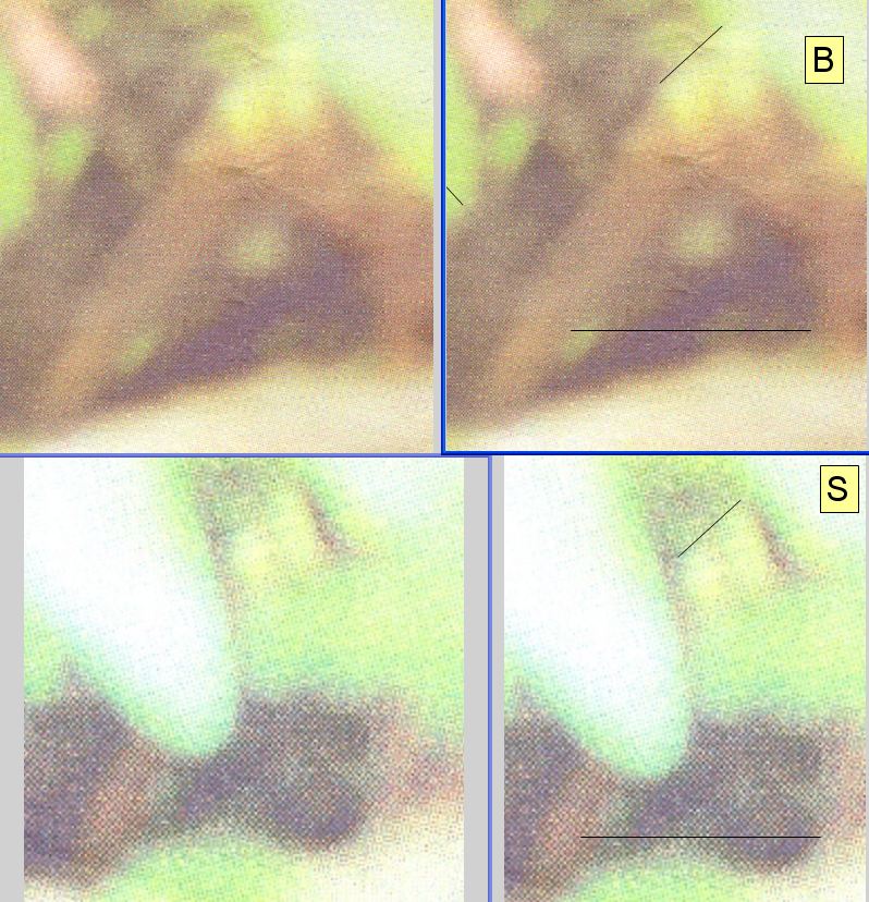
Figure 03 - The rump.
This image shows the rump area of the thylacine in both the bigger (B, top) and smaller (S, bottom) images. Each image of the rump is shown with measurement lines (at right) and without (at left). The first measurement made was of the width of the two stripes. The smaller image was measured, then the line duplicated and placed over the bigger image. This author finds the comparison inconclusive for two reasons; the top of the right-most stripe in image B is partly obscured by a leaf and the distinction between the somewhat falsely coloured stripe and the leaves in the image background is difficult to discern. As such, the accuracy of the placement of the lines is questionable.
The second (horizontal) line was placed at the widest visible point between the tail and hind leg in image B. The line was then copied and aligned with the hind leg in image S. It is clear the extent of the line protrudes over the tail of the animal but no conclusion can be drawn as it cannot be ascertained that the line has been placed at the same vertical distance from some fixed reference point (such as the ground or the rump); this lead to figure 04
Inconclusive
Let me emphasise that I found the analysis involving Figure 03 to be inconclusive. However, this analysis inspired Figure 04 (below). If we can vertically fix the position of the horizontal line in Figure 03, from some known point, then we can ask whether the tail has moved with respect to the rear portion of the hind leg.
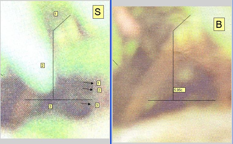
Figure 04 - checking for movement in the tail.
Figure 04 was created to address one shortcoming in figure 03 – namely the unreliability of the horizontal measurement in figure 03. In figure 04 a vertical line was added to image B which joined the end of the rump measurement to the horizontal measurement between the leg and the tail (at its widest visible point in that image). The indicated length of the line (5.95cm) is irrelevant. The same line was copied and duplicated on image S (line 2). Horizontal line 3 was then moved vertically into position so as to touch line 2 in the same way as shown on image B. Line 3 was also moved horizontally into position so as to touch the hind leg in the same way as shown on image B. The horizontal lines are of identical length between images. In image S, the horizontal line (3) clearly protrudes significantly over the tail for more than 50% of its thickness.
Note that for the horizontal positioning of the line, three visible points on the hind leg are indicated by the arrows. The exact point of contact between line 3 and the hind leg is obscured (in image S) by a mark which resembles vegetation, lying between arrows 2 and 3. The arrows provide points of reference either side of the contact point allowing the contact point to be interpolated.
Additional information is yielded by comparing the line subtended by the three arrows in image S (ie the angle of the hind leg) with the angle of the hind leg in image B; the hind leg appears to be “leaning slightly to the right of vertical”; that is, the top of the line is positioned slightly further right than the bottom. Looking at a point on the hind leg in image B which is equivalent in vertical distance from the horizontal line as that shown by arrow 1, clearly shows the leg “curving to the left” as you progress up the leg.
From this figure we can conclude that
- the leg is depicted at a different angle in each image
- and the tail is further from the leg in image B
One explanation for these observations is that in fact the photos depict a real thylacine or a thylacine taxidermy, and the tail moved with respect to the hind leg between photos. In addition or separately, the hind leg changed position with respect to the camera.
However, before a conclusion can be drawn, consideration must be given to alternative explanations. At this point we analyse the possibility that the photograph is of an inanimate object and question how the observed differences between the photos could be achieved by movement of the camera relative to the animal. It is sufficient to exclude all other photographed objects from the analysis as they have no bearing on the way in which the camera perceives the animal. Subsequent analyses will continue to examine the relative positioning of other elements in the photographs with the intent of determining whether or not consistency exists between the image.
(In plain terms, further analysis will continue to investigate whether it’s plausible to assert that the animal moved relative to its environment between photos.)
Copyright notice
The Emmerichs photographs, and derivative works, were removed from the former version of this website (at www.wherelightmeetsdark.com) in 2006 on request of Klaus Emmerichs. They were republished on 12 June 2019 under the defence of Fair Dealing under Australian copyright law. The original images and their elements as incorporated in derivative works are presented as they were published in the print version of Tasmanian newspaper, the Sunday Tasmanian in 2006. An attempt was made to contact Mr Emmerichs on 17 Mar 2017 ahead of the re-publication of these images but he could not be reached. For full details on how WLMD respects and supports copyright, please refer to the Terms and conditions of use. This note added 12 June 2019.
Revisions
12 June 2019 - Republication of images, minor caption changes and copyright notice.